






















In Stock
Intra Oral Dental Camera
Integrated Multi-LED Illumination: Features a high-intensity, shadowless LED array surrounding a precision optical lens to ensure consistent color temperature and visibility in deep posterior regions.
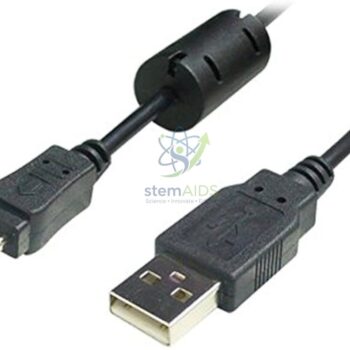
Plug-and-Play USB Connectivity: Engineered with a high-speed digital interface that supports seamless integration with major dental practice management software without requiring proprietary capture cards.
₹2,105.00
Quick Answer: The Intra Oral Dental Camera is a slim handheld wand camera that is placed inside the mouth to show teeth and gums on a computer or monitor. Dental and dental hygiene courses use an intra oral dental camera so that a whole group can see what normally only the person at the chair can see.
The Intra Oral Camera in Dental Teaching
Looking into a mouth is a one-person job: only the operator has a clear view. An intra oral dental camera changes that by carrying a lens and light to the tooth surface and sending the picture to a screen. In a teaching clinic or skills lab, a supervisor can show students a buccal surface, a restoration margin or plaque deposits on a phantom head or a volunteer, and the whole group sees the same close-up.
The camera is also a documentation tool. Still images captured before and after a scaling exercise show students what they achieved, and images can be discussed later without the model or volunteer in the chair. The listing does not state the resolution, connection or software, so check these, along with the computer operating system you use, when enquiring.
This page describes the camera as a teaching and documentation instrument. It is not presented as a diagnostic device, and anyone planning to use it in routine patient care should check the local regulatory requirements for dental imaging equipment and follow the supplied instructions.
Specifications
| Item | Handheld intra oral camera |
| Use | Close-up viewing and image capture inside the mouth |
| Display | External computer or monitor |
| Infection control | Used with a disposable barrier sleeve for each person |
| Resolution, connection and software | Confirm at enquiry |
Applications
- Demonstrating tooth surfaces and restorations to a group in dental or dental hygiene training
- Recording before-and-after images of scaling or polishing exercises on phantom heads
- Showing oral anatomy such as cusps, fissures and the gingival margin at close range
- Building an image bank for tutorials and assessment discussions
Care & Handling
- Cover the intra oral dental camera wand with a new disposable barrier sleeve for every person, and never reuse a sleeve.
- Disinfect the wand between uses with a product the camera’s instructions permit; do not immerse or autoclave it unless the instructions allow it.
- Store the wand in its holder, not loose in a drawer, so the lens window does not get scratched.
- Coil the cable loosely and avoid kinking it where it joins the handpiece.
Why Choose LabEquip
Dental colleges, dental hygiene schools and clinical skills labs buy intra oral cameras to make chairside teaching visible to a group. The camera is listed under Microscope & Optics; describe your display set-up and course needs to LabEquip through the contact page.
Frequently Asked Questions
What does an intra oral camera show that a mouth mirror does not?
A mouth mirror gives the operator alone a reflected view. The camera shows an enlarged image on a screen that several people can watch at once, and it can capture still images for later discussion, which a mirror cannot.
Is the camera a diagnostic device?
It is described here as a teaching and documentation tool. Diagnosis rests with a qualified dentist using appropriate clinical methods, and any clinical use of the camera should follow local regulations for dental equipment.
How is cross-infection prevented?
A new disposable barrier sleeve goes over the wand for each person and is removed after use, followed by surface disinfection with a product approved in the camera’s instructions. The sleeve should sit snugly over the lens window so the image stays clear.
Can it be used on phantom heads and typodonts?
Yes. Phantom heads and typodont models are ideal for practice, because students can learn to position the camera and capture images without time pressure or discomfort for a patient.
Why are the images blurred or too bright?
Blur usually comes from holding the tip too close or moving during capture, so hold it steady at the working distance given in the instructions. Glare from wet enamel is reduced by drying the surface gently with air and angling the camera slightly.
What computer set-up is needed?
That depends on the connection and software supplied with the camera. Check the required operating system and whether a driver is needed, and test the camera on the teaching computer or monitor before the first class.
Last Updated: September 2026
You must be logged in to post a review.
Bestsellers


Sale




Reviews
There are no reviews yet.