













In Stock
Female Half Basin
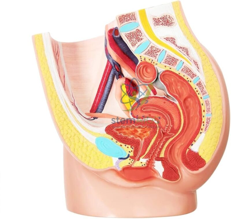
Demi bassin féminin Engineered with a 1:1 anatomical scale providing a detailed medial view of the pelvic cavity, including the internal iliac artery and sacral plexus for advanced vascular and neurological mapping.
Medical-Grade PVC Construction Fabricated using high-durability, non-toxic polymers that resist chemical degradation and maintain color-coded tissue differentiation across high-cadence clinical demonstration cycles.
₹25,430.00
Quick Answer: The Female Half Basin is a female half pelvis section model: the female pelvis cut in the midline so the organs and bones can be seen in section. Basin comes from the French word bassin, meaning pelvis. The model is used in anatomy, nursing and midwifery teaching.
What the Half Pelvis Section Shows
The product photo shows the model cut in the median plane, with the lower lumbar vertebrae, sacrum and pubic bone in section and the pelvic organs between them, in order from front to back: bladder, then uterus and vagina, then rectum, with the abdominal wall, muscle and fat layers coloured to tell them apart. Seeing the organs stacked front to back in their true order is the main advantage of a sectioned model over a whole one.
The section is especially useful for teaching the pelvic floor. The levator ani and related muscles form a sling beneath the organs, with openings for the urethra, vagina and anal canal. Pregnancy, childbirth and ageing can weaken this support, and the model helps explain continence, pelvic floor exercises and why organs can descend in prolapse. The position of the uterus above and behind the bladder also shows why a full bladder is requested for some pelvic ultrasound scans.
The listing does not state the scale, the material, whether any parts are removable or whether a key to the structures is included. LabEquip also lists the human female half pelvis, which covers the same region; ask which suits your course if you are choosing between them.
Specifications
| Model of | Female pelvis in median (midline) section |
| Bones in section, as shown in the product photo | Lower lumbar vertebrae, sacrum and pubic bone |
| Organs | Bladder, urethra, uterus, vagina, rectum |
| Soft tissues | Pelvic floor muscles, abdominal wall and fat layers |
| Scale, material and removable parts | Confirm at enquiry |
Teaching Uses
- Anatomy lessons on the relations of the female pelvic organs
- Midwifery and nursing teaching on the pelvic floor, continence and prolapse
- Preparing students for urinary catheterisation and pelvic examinations
- Explaining the full-bladder technique used in some pelvic ultrasound scans
Care & Handling
- Stand the model on its base or flat cut face and avoid resting it on the painted section surface.
- Clean with a soft, slightly damp cloth; solvents and abrasives damage the colour coding.
- Cover it or keep it in a cupboard between sessions to protect it from dust and sunlight.
Why Choose LabEquip
Nursing schools, midwifery programmes and anatomy departments buy half pelvis models to teach organ relations that are hard to see in a whole-pelvis model. The Female Half Basin is part of LabEquip’s biology lab products; to confirm details, use the contact page.
Frequently Asked Questions
Why is the model called a half basin?
Bassin is the French word for pelvis, and the name has been translated literally. The model is a female half pelvis: the pelvis cut down the midline so that one half shows the internal organs in section.
What is the pelvic floor?
It is a sheet of muscles and connective tissue, mainly the levator ani, stretching across the bottom of the pelvis. It supports the bladder, uterus and rectum and helps control the openings of the urethra and anus.
What is pelvic organ prolapse?
It happens when weakened pelvic floor support lets an organ such as the bladder, uterus or rectum drop down into the vagina. Pregnancy, childbirth, ageing and long-term straining are common contributing factors.
Why is a full bladder needed for some pelvic ultrasound scans?
A full bladder lifts gas-filled bowel out of the way and acts as a clear window through which sound waves reach the uterus and ovaries. The section shows how closely the bladder and uterus lie.
Why does the female urethra matter for infection?
It measures only about 4 cm and its opening lies near the vaginal and anal openings, which gives bacteria a short route into the bladder. Women therefore get urinary tract infections far more often than men.
How does this model differ from a whole female pelvis model?
A whole pelvis model shows the bony ring and its measurements, which matter in childbirth. A half pelvis section shows the organs inside and how they relate to each other, which a closed bony pelvis hides.
Last Updated: September 2026
You must be logged in to post a review.
Bestsellers




Reviews
There are no reviews yet.