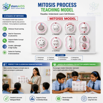
Quick Answer: The Mitosis Process Teaching Model shows the sequential stages of mitotic cell division — prophase, metaphase, anaphase and telophase — so students can trace how a single cell duplicates and divides into two genetically identical daughter cells. LabEquip supplies it as part of the Biology Models range, alongside the Meiosis Visualization Model used for reproductive cell-division teaching.
Product Description
Mitosis is one of the first cell-division processes students study in biology, and it is easier to understand as a sequence of physical stages rather than as a single static diagram. This Mitosis Process Teaching Model from LabEquip is built to lay out the stages of mitotic division so students can trace chromosome behaviour step by step.
Teachers typically use a model like this to walk through prophase (chromosome condensation), metaphase (chromosome alignment), anaphase (chromatid separation) and telophase (nuclear division and cytokinesis), reinforcing why mitosis produces two daughter cells that are genetically identical to the parent cell — unlike meiosis, which produces genetically varied reproductive cells.
What This Model Helps Students Learn
- Identify and sequence the stages of mitosis: prophase, metaphase, anaphase and telophase.
- Trace chromosome behaviour — condensation, alignment, separation and division — through each stage.
- Explain why mitosis produces two genetically identical daughter cells.
- Distinguish mitotic (somatic) cell division from meiotic (reproductive) cell division.
- Connect mitosis to growth, tissue repair and asexual reproduction concepts in the syllabus.
- Practice labelling and describing cell-division stages for practical exams.
Specifications
| Attribute | Detail |
|---|---|
| Product name | Mitosis Process Teaching Model |
| Category | Biology Models – Biology Laboratory Products |
| Product type | Cell-division (mitosis) teaching model |
| Typical use | Teaching mitotic cell-division stages and chromosome behaviour |
| Recommended for | Schools, colleges, biology laboratories, cell-biology educators |
Safety Note
This is a static, non-electrical, non-chemical teaching model intended for normal classroom handling. Handle any detachable stage pieces carefully to avoid breakage, and keep small detachable components away from very young children because of choking-hazard risk. Recommended for use under teacher supervision during classroom demonstrations.
Care & Maintenance
- Keep the model clean and free of dust, wiping with a dry or lightly damp cloth as suited to the material.
- Store away from direct sunlight and excess heat to prevent warping, fading or discolouration.
- Handle any detachable stage pieces gently and store them together, in sequence, so none go missing.
- Check labels and connecting points periodically for damage before each classroom session.
Why Choose LabEquip
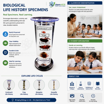
LabEquip supplies biology teaching models, cell-biology teaching aids and laboratory equipment to schools, colleges and technical training institutes across India. Alongside this Mitosis Process Teaching Model, our Biology Models range includes the Meiosis Visualization Model for reproductive cell-division teaching, plus the DNA Double Helix Model and Biological Life History Specimens set. Browse our full Biology Models category for the complete range.
Related Products
- Meiosis Visualization Model: A meiosis model for contrasting the two meiotic divisions and four varied cells with one mitotic division.
- Model of a Cell in Mitosis: A set showing an animal cell at successive mitosis stages, from interphase to two daughter cells.
- Mitosis Manipulatives Kit: Cell, spindle and chromosome pieces that small groups arrange stage by stage to model mitosis.
- DNA Double Helix Model: A DNA double helix model for explaining the copying of DNA during interphase before division.
Frequently Asked Questions
What are the stages of mitosis in the correct order?
Mitosis runs through prophase, metaphase, anaphase and telophase, usually followed by cytokinesis, the division of the cytoplasm. A common memory aid is PMAT. Interphase, when the cell grows and copies its DNA, comes before mitosis and is not itself a stage of mitosis, a point the Mitosis Process Teaching Model helps make clear.
How can you tell metaphase from anaphase on a mitosis model?
In metaphase the chromosomes, each still made of two sister chromatids, line up across the middle of the cell on the equator, attached to spindle fibres. In anaphase the chromatids have been pulled apart and move towards opposite poles, so the model shows two separate groups of V-shaped chromosomes heading away from the centre.
What happens during interphase before a cell divides by mitosis?
Interphase is the longest part of the cell cycle. The cell grows, makes new organelles and copies its DNA, so each chromosome becomes two identical sister chromatids joined at the centromere. Only after this copying can mitosis share an identical set of chromosomes between two new cells.
How is mitosis different in plant cells and animal cells?
The chromosome movements are the same, but the endings differ. An animal cell divides its cytoplasm by a cleavage furrow that pinches the cell in two, and it has centrioles at the spindle poles. A plant cell has a rigid wall, so it builds a cell plate across the middle that becomes the new wall, and it has no centrioles.
Why does mitosis produce two genetically identical cells?
Every chromosome is copied exactly before division, and in anaphase the two copies of each chromosome go to opposite poles. Each daughter cell therefore receives one complete, identical set. That is why mitosis is used for growth, repair and asexual reproduction, while meiosis is needed to make varied gametes.
Where can students see real cells dividing by mitosis?
An onion root tip squash is the classic practical. The tip is softened in acid, stained and pressed under a coverslip, and many cells in different stages can be seen at high power. Students then match what they see to the stages shown on the model and count cells in each stage to estimate the mitotic index.
Last Updated: November 2026