Quick Answer: The Amoeba Cell Anatomy Model is a much-enlarged classroom model of Amoeba, a single-celled protozoan with no fixed shape, used to explain how one cell moves, feeds, removes excess water and reproduces.
Amoeba: Life Without a Fixed Shape
Amoeba is often the first single-celled organism students meet, and its defining feature is that it keeps changing shape. The Amoeba model captures one moment of that movement, so the teacher can explain what is happening inside the cell instead of chasing a live specimen that never holds still.
An Amoeba model is generally used to explain pseudopodia, the temporary extensions of cytoplasm that the cell pushes out to crawl and to surround food. Lessons cover phagocytosis, in which pseudopodia enclose a food particle in a food vacuole for digestion; the single nucleus that controls the cell; and the contractile vacuole, which pumps out excess water taken in from the freshwater surroundings.
The cytoplasm itself is worth explaining. It has a clear, firmer outer layer, the ectoplasm, and a granular, more fluid inner layer, the endoplasm, whose flow into a growing pseudopodium carries the cell forward. Amoeba reproduces by binary fission, the nucleus dividing first and then the cytoplasm.
Specifications
| Organism | Amoeba (amoeboid protozoan) |
| Format | Enlarged classroom model in three dimensions |
| Lesson topics | Movement, phagocytosis, osmoregulation and binary fission |
| Course fit | School biology and first-year college zoology |
Applications
- First lessons on unicellular organisms and the characteristics of life
- Explaining phagocytosis, later linked to white blood cells in immunity lessons
- Comparing amoeboid movement with the ciliary movement of Paramecium
- Asexual reproduction lessons on binary fission
Care & Handling
- Dust the Amoeba model with a soft brush before each term.
- Keep it away from heaters and bright windows to prevent warping and fading.
- Return it to its shelf after the lesson rather than leaving it on a wet practical bench.
Why Choose LabEquip
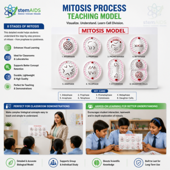
Most schools pair the Amoeba Cell Anatomy Model with the Paramecium Educational Model for a protozoa lesson, and the Mitosis Process Teaching Model supports the nuclear-division step of binary fission. Orders go through the contact page, and all three sit in Biology Models.
Related Products
- Paramecium Educational Model: An enlarged Paramecium for comparing ciliary movement and a fixed oral groove with amoeboid movement and feeding.
- Amoeba Model, Superior: A superior-grade Amoeba proteus model showing pseudopodia, ectoplasm, endoplasm, nucleus and both kinds of vacuole.
- Zoology Prepared Slides: Permanent zoology slides, including whole mounts of small organisms such as Amoeba, for the microscope follow-up.
- Cavity Slide – Single, Double and Triple: Glass slides with ground wells that hold a drop of pond water so live protozoa can be watched.
- Animal Cell Study Model: An enlarged animal cell model for comparing a free-living single cell with a cell from a multicellular body.
Frequently Asked Questions
What is the difference between ectoplasm and endoplasm in an amoeba?
The ectoplasm is the clear, firmer outer layer of cytoplasm just inside the cell membrane, while the endoplasm is the granular, more fluid inner layer that holds the nucleus and vacuoles. On the Amoeba Cell Anatomy Model the two layers help explain movement: endoplasm streams forward into a growing pseudopodium and sets as ectoplasm at its edges.
How do pseudopodia let an amoeba both move and feed?
A pseudopodium is a temporary bulge of cytoplasm pushed out in one direction. When the rest of the cell flows into it, the amoeba creeps forward; when two pseudopodia curve round a food particle and meet, they trap it. The same structure therefore serves for locomotion and for catching food, which is why the model shows the cell mid-movement.
What happens to food after an amoeba engulfs it?
The trapped particle sits inside a food vacuole, a small sac surrounded by membrane. Digestive enzymes enter the vacuole and break the food down, the useful products pass into the cytoplasm, and the undigested remains are expelled at the cell surface wherever the vacuole happens to reach it, since an amoeba has no fixed anal opening.
Why does a freshwater amoeba need a contractile vacuole?
Its cytoplasm is more concentrated than pond water, so water keeps entering the cell by osmosis. The contractile vacuole collects this excess water, swells and then empties it outside, again and again, so the cell does not burst. This is the osmoregulation topic listed for the model, and it makes a good link to water balance in larger animals.
What are the steps of binary fission in an amoeba?
Binary fission begins once the amoeba has grown to full size. The nucleus divides first, then the cytoplasm pinches in the middle until two smaller daughter cells separate, each with its own nucleus. No partner is needed, so it is a form of asexual reproduction, and each daughter amoeba grows and can divide again.
Which level of study is the Amoeba Cell Anatomy Model meant for?
It is listed for school biology and first-year college zoology. Younger classes use it to meet a single-celled organism and the characteristics of life; college students use it for amoeboid movement, phagocytosis and osmoregulation in more detail, and to compare protozoa with other unicellular forms before examining slides.
Last Updated: November 2026