


































In Stock
Inner Ear Model
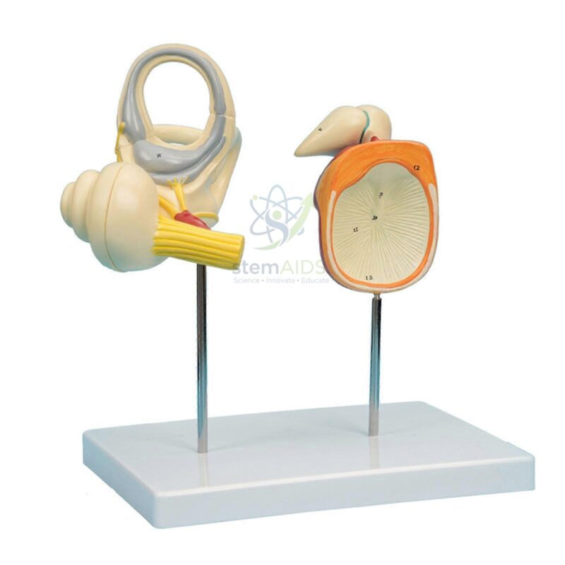
Modèle d'oreille interne Features a 5x life-size representation of the osseous and membranous labyrinths, including the vestibule and the organ of Corti, calibrated for high-precision tactile analysis.
Removable Vestibulocochlear Nerve Engineered with a detachable cranial nerve VIII (auditory-vestibular nerve) to demonstrate the neural pathways responsible for equilibrium and sound transduction in a standardized clinical format.
$139.33
Quick Answer: The Inner Ear Model is an enlarged inner ear model showing the labyrinth: the snail-shaped cochlea for hearing and the vestibule and three semicircular canals for balance, together with the nerve that carries their signals to the brain. It is used in biology, audiology foundation and nursing lessons on hearing and balance.
Hearing and Balance in One Structure
The product photo shows two pieces on metal rods above a white base. One is the labyrinth, with the coiled cochlea, the three looping semicircular canals and a yellow nerve bundle leaving it. The second piece appears to show the eardrum with small bones of the middle ear attached, which helps link sound collection to the inner ear. The listing does not state the magnification or the number of parts, so confirm both before ordering.
The cochlea handles hearing. Vibrations passed from the eardrum through the ossicles push on the oval window and set the fluid inside the cochlea moving. This moves the basilar membrane, which is narrow and stiff near the base of the coil and wider and more flexible toward the tip, so high pitches stimulate hair cells near the base and low pitches those near the apex. The hair cells turn the movement into signals in the cochlear nerve. Hair cells destroyed by prolonged loud noise do not regrow in humans, which is why noise-induced hearing loss is permanent.
The vestibular system handles balance. The three semicircular canals sit roughly at right angles to one another and detect turning of the head in any direction, while the utricle and saccule in the vestibule sense gravity and straight-line acceleration. Conflicting signals from these organs and the eyes explain motion sickness. For the outer and middle ear as well, see the human ear model, 3 parts.
Specifications
| Model of | Enlarged human inner ear (labyrinth) with its nerve |
| Parts in the product photo | Labyrinth, plus a second piece with the eardrum and ossicles, on rods and a base |
| Hearing structure | Cochlea |
| Balance structures | Vestibule and three semicircular canals |
| Magnification and number of parts | Confirm at enquiry |
Where It Fits in the Syllabus
- Biology lessons on the sense organs, hearing and balance
- Explaining how pitch is detected along the length of the cochlea
- Health education on noise exposure and hearing protection
- Nursing and audiology foundation teaching on vertigo, motion sickness and ear examination
Care & Handling
- Lift the inner ear model by its base; the pieces sit on thin rods that bend if the model is carried by the labyrinth.
- Take care with the semicircular canals, which are the thinnest parts and snap under sideways pressure.
- Dust with a soft brush and keep the model covered between lessons.
Why Choose LabEquip
Biology departments, nursing schools and audiology foundation courses buy an enlarged inner ear because the real structure is tiny and buried in the temporal bone. LabEquip lists it under biology lab products; send questions on magnification through the contact page.
Frequently Asked Questions
What is the difference between the inner, middle and outer ear?
The outer ear collects sound and channels it to the eardrum. The middle ear is an air-filled space where three small bones pass vibrations inward. The inner ear is a fluid-filled labyrinth containing the organs of hearing and balance.
How does the cochlea tell high notes from low ones?
The basilar membrane inside the cochlea changes in width and stiffness along its length. High-pitched sounds make it vibrate most near the base of the coil and low-pitched sounds near the tip, so different hair cells respond.
What do the semicircular canals do?
They detect rotation of the head. Each canal is filled with fluid that lags behind when the head turns, bending hair cells in a swelling at its base. With three canals set at different angles, turning in any direction is sensed.
Why does spinning make you dizzy?
During a long spin the fluid in the canals starts moving with the head. When you stop, it keeps moving for a moment, so the canals signal turning while the eyes report that you are still, and the mismatch is felt as dizziness.
Why is noise-induced hearing loss permanent?
Loud noise damages the delicate hair cells of the cochlea, and in humans these cells do not regrow. Limiting exposure time and wearing ear protection in noisy places helps prevent the damage.
Does this model show the middle ear bones?
The product photo shows a second piece that appears to be the eardrum with ossicles attached, next to the labyrinth. The exact parts supplied should be confirmed, and a full ear model shows the outer, middle and inner ear together.
Last Updated: September 2026
You must be logged in to post a review.
Bestsellers




Reviews
There are no reviews yet.