











In Stock
Male Basin Model
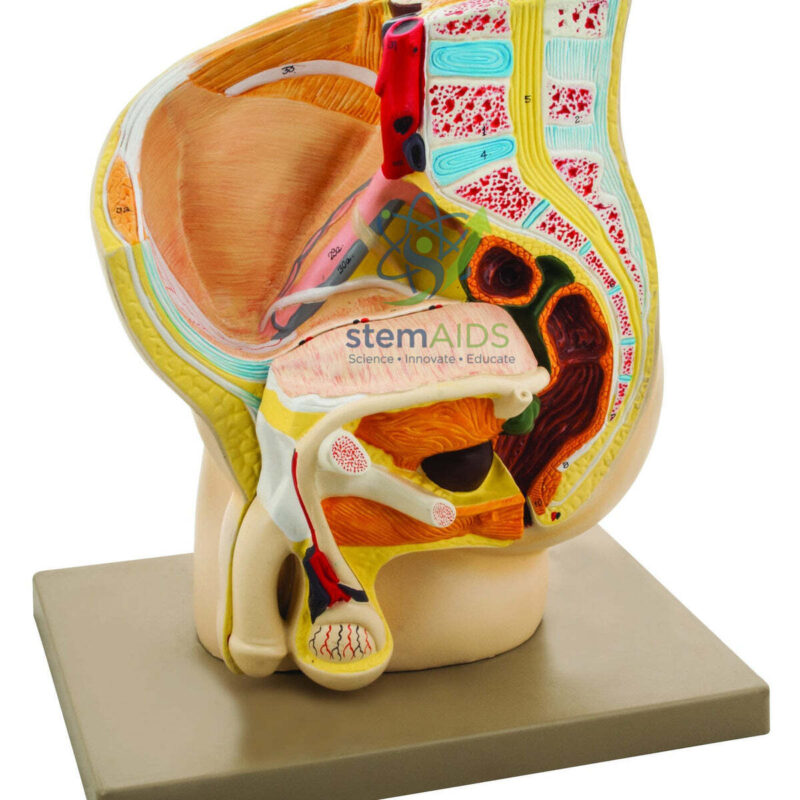
Modèle de bassin masculin Features a 1:1 scale medial section providing detailed topological views of the pelvic floor, seminal vesicles, and testicular morphology for advanced physiological study.
High-Density Bio-Polymer Matrix Fabricated from medical-grade, non-toxic PVC that maintains color-coded tissue differentiation and tactile integrity through high-frequency tactile examination and repetitive institutional use.
$265.13
Quick Answer: The Male Basin Model is a male pelvis section model, cut in the midline and mounted on a base, that shows the bladder, prostate, urethra, penis, testis and rectum in their natural positions. It is used in biology, anatomy and nursing lessons on the male reproductive and urinary tracts; “basin” here is a literal translation of bassin, the French word for pelvis.
Following the Male Reproductive Tract in Section
The product photo shows the model cut down the median plane, with the lower spine and sacrum behind, the pubic bone in front and the organs lying between them. Unlike many half-pelvis models, this one continues below the pelvic floor to show the penis and a testis within the scrotum in section, so a teacher can walk through the whole route of sperm on a single piece: from the testis to the epididymis on its back surface, up the ductus deferens, past the seminal vesicle behind the bladder, through the ejaculatory duct inside the prostate and out along the urethra.
The section also shows how the urinary and reproductive tracts come to share one channel. The prostate surrounds the urethra just below the bladder neck, and the erectile tissue of the penis, the corpora cavernosa above and the corpus spongiosum around the urethra, can be pointed out on the cut surface. Behind the bladder the rectum lies close to the prostate and seminal vesicles, separated from them by only a thin layer of fascia.
LabEquip also lists a human male half pelvis model of the same region, which suits lessons centred on the urethra and catheterisation. This page describes the Male Basin Model from its product photo; the listing does not state its scale, its material or whether any organs lift out.
Specifications
| Model of | Male pelvis in median (midline) section, on a base |
| Reproductive structures in section | Testis, penis with erectile tissue, prostate |
| Urinary structures | Bladder and urethra |
| Other structures | Rectum, lower spine, sacrum and pubic bone |
| Name | “Basin” is a literal translation of the French bassin (pelvis) |
| Scale, material and removable parts | Confirm at enquiry |
Teaching Uses
- Biology lessons on the male reproductive system and the route taken by sperm
- Health education on testicular self-examination, showing where the testis and epididymis lie
- Explaining vasectomy and male contraception with reference to the ductus deferens
- Nursing and anatomy classes comparing male and female organ arrangement alongside the Woman’s Half Basin
Care & Handling
- Lift the Male Basin Model by its base; the section face and thin projecting parts such as the penis and scrotum are the easiest to chip.
- Dust with a soft brush and wipe marks with a cloth barely dampened with water, since solvents dull the colour coding that separates the organs.
- Store it covered and out of direct sunlight, which fades painted surfaces over time.
Why Choose LabEquip
Schools teaching reproduction, nursing colleges and health-education programmes buy a sectioned male pelvis so learners can see organs that diagrams flatten. The model belongs to LabEquip’s biology lab products range; to ask about scale or about pairing it with a female section, write through the contact page.
Frequently Asked Questions
Why is it called a male basin model?
Bassin is the French word for pelvis, and the listing has translated it literally. The item is a model of the male pelvis cut in the midline, showing the pelvic organs and external genitalia in section.
What route do sperm take through the structures on this model?
Sperm are made in the testis and mature in the epididymis. They travel up the ductus deferens, which joins the duct of the seminal vesicle to form the ejaculatory duct inside the prostate, and then leave through the urethra in the penis.
How does a male pelvis section differ from a female one?
There is no uterus or vagina, so the bladder lies directly in front of the rectum. The prostate sits below the bladder around the urethra, and the male urethra is much longer because it runs through the penis.
Can the model help with teaching testicular self-examination?
Yes. It shows where the testis and epididymis lie, and learners often mistake the soft epididymis at the back of the testis for a lump. Any new lump, swelling or change in a testis should be checked by a doctor.
What is cut in a vasectomy?
The ductus deferens on each side is cut or sealed in the scrotum, so sperm can no longer reach the urethra. Most of the fluid in semen comes from the seminal vesicles and prostate, so its volume changes very little.
How should the section surfaces be cleaned?
Use a soft brush for dust and a cloth lightly dampened with water for marks. Avoid alcohol, solvents and abrasive cleaners, which can remove the paint that distinguishes the organs.
Last Updated: September 2026
You must be logged in to post a review.
Bestsellers




Reviews
There are no reviews yet.