
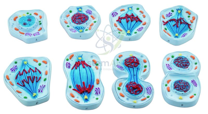
Quick Answer: The Model of a Cell in Mitosis is a mitosis model that shows an animal cell at successive stages of division, from interphase through prophase, metaphase, anaphase and telophase to the two daughter cells formed by cytokinesis. Biology teachers use it to show how copied chromosomes are shared equally between new cells.
Reading the Stages on the Mitosis Model
The product photo shows the model as a series of eight separate cell pieces laid out in order, each with coloured chromosomes, spindle fibres and organelles. The cells have no cell wall, so they represent animal cells.
In interphase the chromosomes are not visible as separate threads, because the DNA is spread through the nucleus while it is copied. In prophase each chromosome condenses into two sister chromatids joined at a centromere, and the centrosomes move apart as the spindle forms. Once the nuclear envelope has broken down, the chromosomes line up across the middle of the cell at metaphase. At anaphase the sister chromatids are pulled to opposite poles, and at telophase new nuclear envelopes form around each set while a cleavage furrow begins to pinch the cytoplasm in two.
Laying the pieces side by side makes testing easy: shuffle them and ask for the correct order, or point to one piece and ask what has just happened and what comes next. For comparing mitosis with the reduction division that makes gametes, the animal meiosis model follows the same style of teaching.
Classroom Uses
- Sequencing exercises in which students arrange the stages of mitosis in order
- Explaining why each daughter cell receives an identical set of chromosomes
- Contrasting cytokinesis in animal cells with the cell plate formed in plant cells
- Revision before onion root tip practicals, linking model stages to what is seen under the microscope
Care & Handling
- Keep the mitosis model pieces in a divided tray or box in stage order, so a missing piece is noticed at once.
- Handle the pieces by their edges; the raised chromosomes and spindle fibres are the parts most likely to snap.
- Wipe with a soft, dry or barely damp cloth, never with solvents that lift the colours used to tell chromosomes apart.
Specifications
| Model of | Animal cell at successive stages of mitosis |
| Pieces in the product photo | Eight cells, from interphase to two daughter cells |
| Features | Chromosomes, spindle fibres, centrosomes, nucleus and cytoplasmic organelles |
| Cell type | Animal cell: no cell wall, divides by a cleavage furrow |
Why Choose LabEquip
Secondary schools and pre-university biology departments buy stage-by-stage mitosis models because students can handle the sequence rather than memorise it from a page. The set is part of LabEquip’s biology lab products; ask about the piece count and size through the contact page.
Related Products
- Mitosis Manipulatives Kit: A manipulatives kit in which students move chromosome pieces through the stages themselves.
- Mitosis Process Teaching Model: A further mitosis teaching model of the stages from prophase to telophase for comparing designs.
- Animal Cell Model: An animal cell model showing the organelles of a non-dividing cell, the starting point before interphase.
- Plant Cell Model, Big: A large plant cell model with a cell wall, for explaining why plant cells divide with a cell plate.
Frequently Asked Questions
What stages are shown on the model of a cell in mitosis?
The product photo shows eight separate animal cell pieces laid out in order: interphase, the stages of prophase, metaphase, anaphase and telophase, and finally two daughter cells formed by cytokinesis. Each piece shows chromosomes, spindle fibres, centrosomes and organelles at that moment of division.
What is the difference between a chromosome and a chromatid on a mitosis model?
Before division each chromosome is copied, so in prophase and metaphase it appears as two identical sister chromatids joined at the centromere. In anaphase the chromatids are pulled apart, and each one is then counted as a separate chromosome in the daughter cell.
How does mitosis in an animal cell end differently from a plant cell?
An animal cell has no cell wall, so in cytokinesis the membrane pinches inwards to form a cleavage furrow that splits the cell in two, as the last pieces of this model show. A plant cell builds a cell plate across the middle from the inside, which becomes the new wall.
What are the spindle fibres and centrosomes doing in each stage?
Centrosomes move to opposite poles in prophase and organise spindle fibres made of microtubules. In metaphase the fibres attach to each chromosome at the centromere and line it up on the equator; in anaphase they shorten and pull the chromatids to the poles, then break down in telophase.
How can students use the mitosis model with a microscope practical?
Use the eight pieces as a reference card set: students identify the stage on each piece, then find matching cells in a stained onion root tip squash or prepared slide. The model shows the stages more clearly, while real cells show how varied and fast-changing they look.
Is a fixed mitosis model or a manipulatives kit better for teaching?
A fixed series of cells is quick to set out, shows every structure clearly and suits demonstration and revision. A manipulatives kit makes students move chromosomes themselves and reveals misunderstandings. Many teachers show the fixed model first and then let groups rebuild the stages with manipulatives.
Last Updated: November 2026







Reviews
There are no reviews yet.