











In Stock
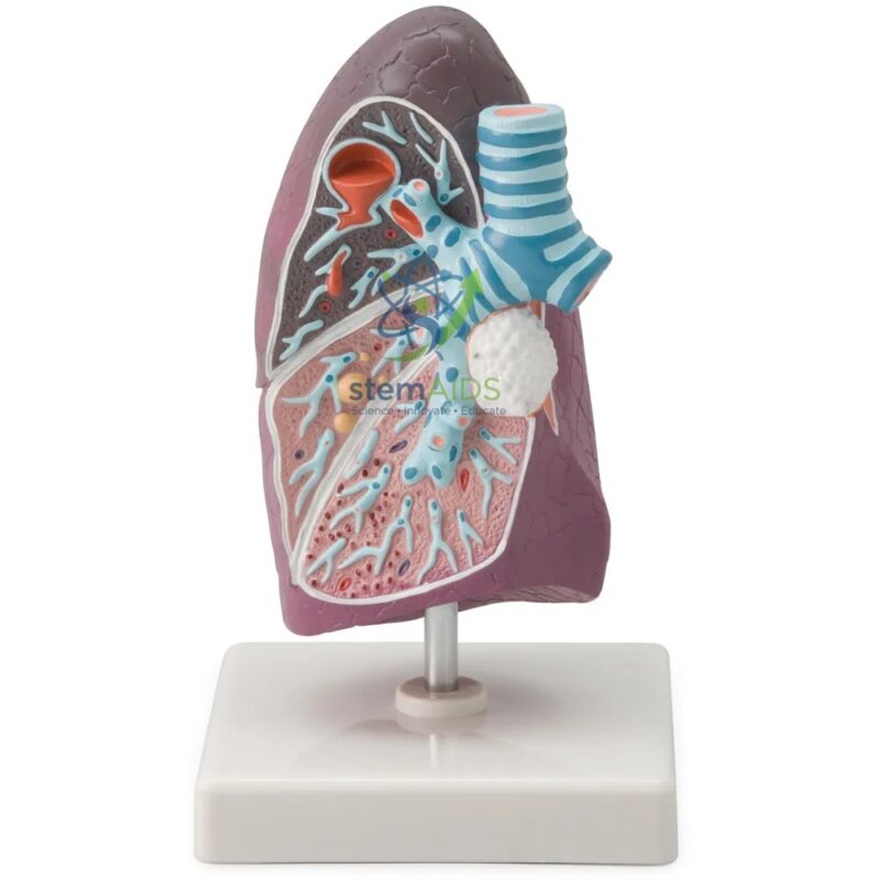
Anatomical Model of a Pathological Human Lung
Modèle anatomique du poumon humain pathologique Features a dual-structure design that contrastively illustrates a healthy left lung against a right lung presenting stage-specific pathologies, including anthracosis, squamous cell carcinoma, and bullous emphysema.
High-Fidelity Histological Texture utilizes advanced medical-grade PVC with hand-painted vascular and bronchial details, providing a tactile, anatomically accurate representation of pleural thickening and neoplastic tissue infiltration.
₹2,105.00
Quick Answer: The Anatomical Model of a Pathological Human Lung is a pathological lung model: a model of a human lung that shows diseased tissue rather than a healthy organ. It is used in biology, nursing and health-education teaching to show how conditions such as smoking-related disease change the structure of the lungs.
Diseased Tissue on a Pathological Lung Model
Most lung models show healthy anatomy: lobes, fissures, the bronchial tree and the pulmonary vessels. A pathological lung model adds the changes that disease causes, so a learner can see what damaged lungs actually look like. The listing does not name the conditions shown, and models of this type differ: some show a single disease, others show several changes on one organ. Confirm which conditions are represented before building a lesson around it.
The changes most often taught with such models are those linked to smoking and polluted air. In emphysema, the walls between alveoli break down into larger air spaces, cutting the surface available for gas exchange and trapping air. Chronic bronchitis thickens the airway lining and increases mucus. Lung cancer usually appears as a tumour mass, often arising from a bronchus near the root of the lung, and may spread to nearby lymph nodes. Dust diseases leave dark deposits in the tissue.
Placed next to a healthy human lung model, the pathological lung helps learners connect symptoms such as breathlessness and chronic cough with physical damage. For the microscopic side of emphysema, a magnified alveoli model shows the air sacs whose walls the disease destroys.
Where It Is Used
- Anti-smoking and health-promotion talks in schools and community programmes
- Nursing and respiratory-care training on COPD and lung cancer
- Biology lessons comparing healthy and diseased gas-exchange surfaces
- Clinic-based patient education, to explain a diagnosis and why stopping smoking helps
Specifications
| Model of | Human lung showing pathological (diseased) changes |
| Anatomy for reference | Lobes, bronchi, pulmonary vessels and root of the lung, as usually shown |
| Typical teaching topics | Smoking-related disease, emphysema, chronic bronchitis, lung cancer |
| Users | Biology, nursing, respiratory-care and health-education classes |
| Conditions shown, scale and mounting | Confirm at enquiry |
Care & Handling
- Lift the pathological lung model by its stand or base, not by the lung, whose thin edges and bronchial parts can chip.
- Dust with a soft brush and wipe with a slightly damp cloth; keep solvents away from the painted diseased areas.
- Store it away from direct sunlight, which can fade the colours that separate healthy and diseased tissue.
Why Choose LabEquip
Health educators and nursing tutors often use this model with a healthy lung so the contrast does the explaining. LabEquip lists it with its biology lab products; for class quantities or to confirm the conditions shown, use the contact page.
Frequently Asked Questions
What does emphysema look like on a lung model?
Emphysema is shown as enlarged, irregular air spaces where the thin walls between alveoli have broken down, while healthy tissue looks fine and spongy by comparison. Losing those walls reduces the area for gas exchange, which is why people with emphysema become breathless.
What is COPD?
COPD, chronic obstructive pulmonary disease, is the umbrella term for long-term lung disease that narrows the airways and limits airflow, mainly emphysema and chronic bronchitis. Smoking is the main cause, and smoke from cooking fuels and workplace dust also contribute.
How does lung cancer usually appear on a model?
It is usually shown as a pale or irregular tumour mass in the lung tissue or around a main bronchus, sometimes with enlarged lymph nodes near the root of the lung. The exact presentation depends on the model.
Why is a pathological model useful for anti-smoking education?
It gives a concrete picture of harm that words alone do not. Showing damaged tissue beside a healthy lung, and linking each change to symptoms such as coughing and breathlessness, makes the risks of smoking easier for students and patients to grasp.
Do the lungs recover after someone stops smoking?
Some things improve: coughing and airway irritation often ease, and the yearly loss of lung function slows towards that of a non-smoker. Alveolar walls destroyed by emphysema do not grow back, which is why stopping early matters.
How should the lung model be cleaned?
Use a soft brush for dust and a cloth dampened with water or mild soap, then dry it. Avoid alcohol, solvents and abrasive pads, which can remove the painted detail that marks the diseased areas.
Last Updated: September 2026
You must be logged in to post a review.
Bestsellers





Reviews
There are no reviews yet.