


Sale
In Stock
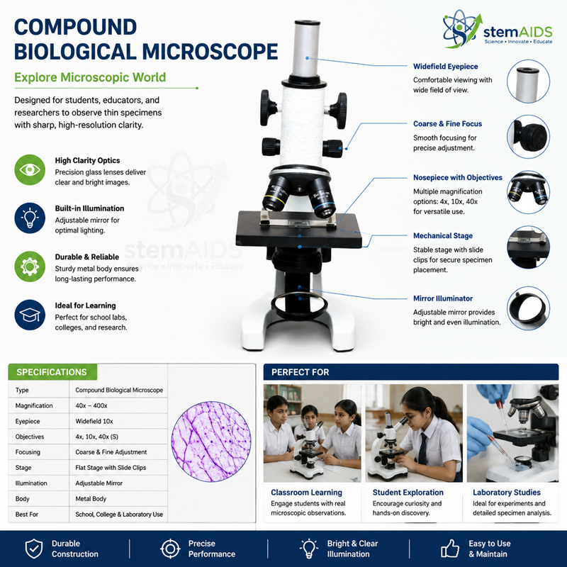
Compound Biological Microscope
Original price was: ₹5,000.00.₹2,490.00Current price is: ₹2,490.00.
Quick Answer: The Compound Biological Microscope is a compound microscope for transmitted-light work, magnifying thin specimens on glass slides through an objective and an eyepiece so that cells, tissues and microorganisms can be studied. Listed without a Student label, it is aimed at general slide work in college and teaching laboratories as well as schools.
Slide Work in the Biology Lab
Most of what biology students examine under a microscope is transmitted-light work: light from below passes through a stained section, smear or wet mount, and the objective collects it. That is the job of a compound biological microscope, as distinct from a dissecting microscope for whole objects or a metallurgical microscope that lights opaque samples from above.
Three controls matter most once the slide is in place. The coarse and fine focus bring the specimen into sharp view. The condenser, where fitted, gathers light onto the specimen and sits close under the slide for high power. The iris diaphragm controls contrast: closing it a little makes unstained cells visible, while opening it gives the resolution needed at high power. Learning these adjustments is what separates a good image from a merely bright one.
The listing does not state the head type, objectives, eyepieces or light source, so check them against the planned work, especially whether a 100x oil-immersion objective is needed for bacteria and blood films. For school class sets, the Student Compound Biological Microscope is the student-labelled model; for image capture, see the Digital Trinocular Microscope with 2MP Camera.
Applications
- Histology practicals on stained sections of animal and plant tissues
- Gram-stained bacterial smears under oil immersion, where an oil objective is fitted
- Haematology teaching with stained blood films
- Botany practicals on hand-cut sections and pollen
Specifications
| Instrument | Compound biological microscope |
| Illumination type | Transmitted light from below the stage |
| Specimens | Stained sections, smears, wet mounts |
| Key controls | Coarse and fine focus; condenser and iris diaphragm where fitted |
| Head, objectives and light source | Confirm at enquiry |
Care & Handling
- Wipe immersion oil off the oil objective after each session with lens tissue, and keep oil away from the dry objectives.
- Lower the stage fully and turn the lowest-power objective into place before storing.
- Turn the brightness down before switching the lamp on or off.
- Store the compound microscope under a dust cover in a dry room, since humidity encourages fungus on lenses and prisms.
Why Choose LabEquip
College biology and microbiology departments, nursing and paramedical training labs and senior school labs buy compound biological microscopes for routine slide work. LabEquip lists it in the Microscope & Optics range; state the objectives and quantity you need on the contact page.
Frequently Asked Questions
What is a compound microscope?
A microscope that magnifies in two stages: the objective forms a magnified image and the eyepiece enlarges it again. Total magnification is objective power multiplied by eyepiece power, and resolving power depends mainly on the objective.
Do I need an oil-immersion objective?
For bacteria, fine blood cell detail and malaria parasites, yes; a 100x oil-immersion objective is the usual tool for that work. For plant and animal tissues, wet mounts and most school practicals, dry objectives are enough. Confirm the objective set at enquiry.
How is immersion oil used?
Focus at high dry power first, swing that objective aside, place a small drop of oil on the slide, then turn the oil objective into the drop and use only the fine focus. Clean the oil off the objective and the slide afterwards.
What does the condenser do?
It concentrates light onto the specimen so the objective receives an even cone of light. Keep it raised close under the slide for high power; lowering it dims the image but can add contrast at low power.
Why are unstained cells hard to see?
They are almost transparent, so they differ little from the background. Close the iris diaphragm partly to raise contrast, or use a stain such as methylene blue or iodine. For live unstained cells, phase contrast gives a clearer image.
How can lens fungus be prevented?
Keep microscopes in a dry, ventilated room under dust covers rather than in closed, damp cupboards. In humid climates store them with a desiccant, and have hazy lenses cleaned by a technician.
Last Updated: September 2026
Bestsellers



