











In Stock
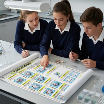
Poster Animal and Plant Cell
Poster Cellule animale et végétale, UV-Stabilized Colorimetric Coding Facilitates rapid student Identification of cellular membranes, organelles, and genetic material storage sites, significantly reducing cognitive load during observational testing.
Standardized Cytological Nomenclature Comparison facile à réaliser, Ensures the accurate Application of scientific terminology in laboratory reports and research papers, aligning with peer-reviewed academic standards.
₹650.00
Quick Answer: The Poster Animal and Plant Cell is a biology wall poster that shows a typical animal cell and a typical plant cell side by side, labelling their organelles so that students can compare the two. It is used in lessons on cell structure and microscopy.
Two Cells Side by Side
Cell structure is usually taught by comparison, and an animal and plant cell poster is built for exactly that. Both cells share a nucleus holding DNA, cytoplasm, a cell membrane, mitochondria where respiration releases energy, ribosomes where proteins are made, and internal membranes such as the endoplasmic reticulum and Golgi apparatus that process and transport materials. The plant cell adds three structures students must name: a cellulose cell wall, chloroplasts for photosynthesis and a large permanent vacuole filled with cell sap.
Posters of this kind are drawn at the level of the electron microscope, showing organelles in more detail than students will ever see through a school light microscope. That gap is worth using: after looking at onion epidermis or cheek cells under the microscope, students compare what they saw (wall, nucleus, perhaps a vacuole) with the poster and learn why the other organelles need far higher magnification.
Shape helps too. Plant cells often look regular and box-like because of the rigid wall, while animal cells are rounder and irregular, a difference the side-by-side drawings make easy to spot.
Specifications
| Subject | Structure of animal and plant cells |
| Organelles typically labelled | Nucleus, cytoplasm, cell membrane, mitochondria, ribosomes, endoplasmic reticulum, Golgi apparatus |
| Plant-only structures | Cell wall, chloroplasts, large permanent vacuole |
| Level of detail | Diagrams at electron-microscope level |
| Level | Middle school to senior secondary biology |
| Size and finish | Confirm at enquiry |
Applications
- The first lesson on cells, before students prepare an onion epidermis slide
- Comparing microscope observations with the labelled diagram
- Revising organelle functions with a matching game using cut-out labels
- Explaining why plants can photosynthesise and animals cannot
Care & Handling
- Hang the poster near the microscopes so students can look up from the eyepiece to the diagram.
- Keep stain bottles such as iodine and methylene blue away from it; splashes leave permanent marks.
- A clip frame lets you put the poster up for the cells unit and protect it for the rest of the year.
Why Choose LabEquip
Biology teachers use this poster alongside ready-made slides from the Prepared Slides range, so students move from diagram to real cells in one lesson. It belongs to the Educational Charts range, where the Virus Chart covers non-cellular life, and class orders can go through the LabEquip contact page.
Frequently Asked Questions
What are the main differences between animal and plant cells?
Plant cells have a cellulose cell wall, chloroplasts and a large permanent vacuole; animal cells have none of these. Animal cells are usually smaller and more irregular in shape, and they contain centrioles, which most plant cells lack.
Which organelles do both cells share?
Both have a nucleus, cytoplasm, cell membrane, mitochondria, ribosomes, endoplasmic reticulum and Golgi apparatus. These carry out tasks every eukaryotic cell needs, such as respiration and protein synthesis.
Why do plant cells need a cell wall?
The cellulose wall supports the cell and stops it bursting when water enters by osmosis. Pressure from the vacuole against the wall keeps plant tissues firm, which is why a watered plant stands upright.
Can students see mitochondria with a school microscope?
Generally not in any detail. A light microscope clearly shows the cell wall, nucleus and sometimes chloroplasts or the vacuole, but mitochondria, ribosomes and the Golgi apparatus need an electron microscope to be seen properly.
Which cells are good for a first microscope practical?
Onion epidermis stained with iodine shows plant cell walls and nuclei clearly, and cheek cells stained with methylene blue show animal cells. Comparing the two with the poster makes the differences concrete.
Do all plant cells have chloroplasts?
No. Root cells and the cells of an onion bulb grow away from light and have no chloroplasts. Cell diagrams usually show a leaf-type cell so that every structure a plant cell can have is included.
Last Updated: September 2026
You must be logged in to post a review.
Bestsellers


Sale


Sale


Reviews
There are no reviews yet.