

In Stock
Skin Cancer Model
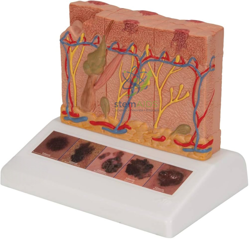
Modèle de cancer de la peau Features a high-resolution cross-section of the epidermis, dermis, and subcutaneous tissue, incorporating six distinct zones that demonstrate the progression of malignant tissue from the stratum basale.
ABCDE Diagnostic Topography Engineered with microscopic surface textures and pigmented irregularities to represent specific clinical indicators of melanoma, including asymmetry, border irregularity, and diameter variation for advanced dermatological training.
₹14,560.00
Quick Answer: The Skin Cancer Model is an enlarged block of human skin in section showing a pigmented tumour growing down from the surface into the deeper layers, with examples of skin lesions pictured on its base. It is used in biology, nursing and health education to teach how skin cancers develop and how suspicious moles are recognised.
How the Skin Cancer Model Is Laid Out
The product photo shows a block of skin with its layers coloured separately: the epidermis on top, the dermis with hair follicles, glands, blood vessels and nerves beneath it, and fatty subcutaneous tissue at the bottom. A dark, pigmented growth extends from the surface down into the dermis, showing a tumour invading deeper tissue. Along the front of the base runs a row of five pictures of pigmented skin lesions. The listing does not name the tumour types represented or say what the pictures show, so confirm these details if the model is chosen for a specific syllabus.
The three main types of skin cancer arise from different cells of the epidermis. Basal cell carcinoma, the most common, comes from the basal layer, grows slowly and rarely spreads. Squamous cell carcinoma arises from keratinocytes higher in the epidermis, often on sun-exposed skin. Melanoma develops from melanocytes, the pigment cells of the basal layer, and is the most dangerous because it can spread through lymph and blood vessels once it grows down into the dermis. How deep a melanoma has grown, measured as the Breslow thickness, is one of the main factors in how it is treated.
Ultraviolet radiation from sunlight and sunbeds is the main avoidable cause, which is why the model is often used in sun-safety teaching. The human skin model shows healthy skin structure for comparison, and the UV A and UV B Sensitive Dosimeter Badge lets students see where and when UV exposure is strongest.
Specifications
| Model of | Enlarged block of human skin with a pigmented tumour |
| Layers in the product photo | Epidermis, dermis, subcutaneous tissue |
| Other structures | Hair follicles, glands, blood vessels and nerves |
| Base | Row of five pictured pigmented skin lesions |
| Tumour types represented and scale | Confirm at enquiry |
Teaching Applications
- Health education on sun safety and the risks of sunbeds
- Teaching the ABCDE signs used to check moles
- Nursing and pharmacy lessons on the types of skin cancer and why depth of invasion matters
- Biology lessons linking the layers of the skin to where different cancers begin
Care & Handling
- Carry the skin cancer model by its base; raised hair shafts and vessel ends on the top and cut faces break off easily.
- Wipe the block and the pictured lesions with a soft, slightly damp cloth, without rubbing the printed pictures.
- Store it covered to keep dust out of the textured surfaces.
Why Choose LabEquip
Nursing colleges, pharmacy courses and school health programmes use a skin cancer model because it makes an unseen process visible: a mole growing downward into the skin. LabEquip lists it with its biology lab products; for scale and content, use the contact page.
Frequently Asked Questions
What are the main types of skin cancer?
Basal cell carcinoma and squamous cell carcinoma, often grouped as non-melanoma skin cancers, and melanoma. They arise from different cells of the epidermis and differ in how quickly they grow and how likely they are to spread.
What is the ABCDE rule?
It is a checklist for moles: Asymmetry, irregular Border, uneven Colour, Diameter larger than about 6 mm, and Evolving, meaning changing in size, shape or colour. A mole showing any of these signs should be checked by a doctor.
Why is melanoma more dangerous than other skin cancers?
Melanoma starts in pigment cells and can grow down into the dermis, where blood and lymph vessels allow it to spread to other organs. Found early, while it is still thin, it is usually treatable by removing it.
What is Breslow thickness?
It is the depth of a melanoma measured in millimetres from the top of the epidermis to the deepest tumour cells. A thicker melanoma is more likely to have spread, so the measurement guides treatment.
How does ultraviolet light cause skin cancer?
UV radiation damages the DNA in skin cells. Repeated damage, especially from sunburn, can lead to mutations that let cells grow out of control. Shade, protective clothing, hats and sunscreen reduce exposure.
Can this model be used to teach self-examination?
Yes. The pictured lesions on the base help learners discuss what to look for in a mole, and the block shows why a lesion that grows or changes needs prompt attention before it grows deeper.
Last Updated: September 2026
You must be logged in to post a review.
Bestsellers





Reviews
There are no reviews yet.