
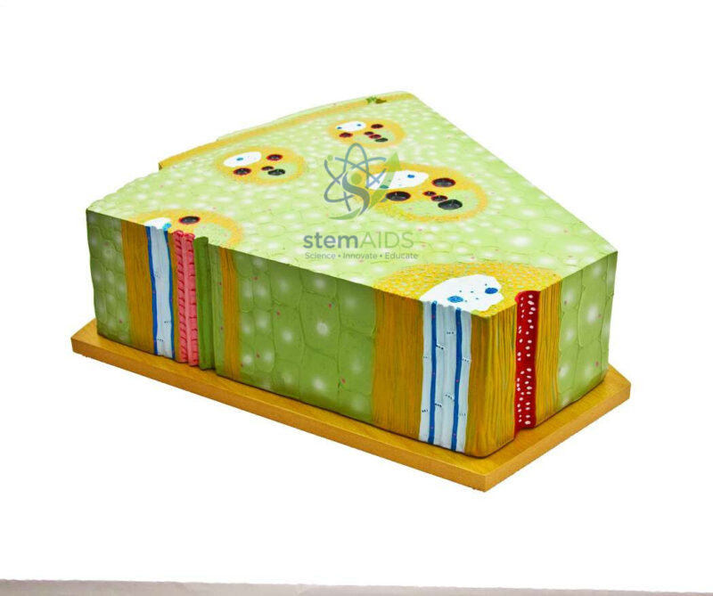
Quick Answer: The Monocotyledon Stem Model is an enlarged teaching model of the internal structure of a monocot stem. The listing photograph shows a block-style piece on a wooden base, with the top face cut as a transverse section and the sides showing the tissues running lengthwise.
Seeing the Monocot Stem in Three Dimensions
Most students first meet the monocot stem as a flat circle on a textbook page: many small vascular bundles scattered through soft ground tissue, with no ring and no clear pith. A block model adds depth. On the top face of the Monocotyledon Stem Model the bundles appear as the familiar scattered spots; on the side faces the same bundles run as long, parallel strands up the stem. Seeing both views together makes it clear that each spot in a cross-section is a strand cut across, not an isolated island of tissue.
The features to point out start at the outside. A single-layered epidermis is covered by cuticle, and under it lies a band of thick-walled supporting cells. Inside is ground tissue made of large, thin-walled parenchyma cells. The bundles are smaller and crowded near the edge and larger and more widely spaced towards the centre. In each one the xylem faces inwards and the phloem outwards, a sheath of fibres surrounds the whole bundle, and no cambium lies between xylem and phloem. That missing cambium explains why most monocot stems do not grow thicker year after year the way a tree trunk does.
Many teaching models follow the maize stem, where a large air or water space appears in older bundles. LabEquip also lists a flat T.S. monocot stem model that shows the cross-section alone.
Specifications
| Model of | Internal structure of a monocotyledon stem |
| Format | Block section on a wooden base, per the listing photograph |
| Views | Transverse face on top; lengthwise tissues on the sides |
| Key features | Epidermis, supporting layer, ground tissue, scattered closed vascular bundles |
Care & Handling
- Lift the model by its base with both hands; painted edges of the block chip if it is knocked against a bench.
- Dust with a soft brush or dry cloth, and avoid solvents that can lift the paint and labels.
- Store away from direct sunlight, which fades the colours used to separate tissue types.
Classroom Uses
- Comparing monocot and dicot stems side by side before students draw labelled diagrams
- Explaining why most monocots lack secondary thickening
- Preparing students to identify bundles in a stained section of maize stem under the microscope
- Revision of plant tissue names ahead of practical examinations
Why Choose LabEquip
Botany teachers usually buy a monocot stem model alongside its dicot counterpart for comparison lessons. LabEquip lists this block model in Biology Lab Products with the matching monocot stem chart. Use the LabEquip contact page to confirm size and base before ordering.
Related Products
- Dicotyledon Stem Model: The dicot stem model shows a ring of open bundles with cambium, the direct comparison for the scattered closed bundles of the monocot stem.
- Model, T. S. Monocot Stem: A flat transverse-section model of a monocot stem that adds the Y-shaped xylem and protoxylem cavity detail seen in maize.
- Monocotyledon Root Model: Shows the monocot root with its endodermis and many alternating xylem and phloem groups, for comparing root and stem anatomy.
- Transverse Section of Monocot Stem Chart: A wall chart of the monocot stem section with labelled bundle sheath, phloem and xylem, useful for whole-class labelling.
Frequently Asked Questions
What does the Monocotyledon Stem Model show on its top and side faces?
The model is a block section on a wooden base. Its top face is cut as a transverse section showing the epidermis, a supporting layer, ground tissue and scattered closed vascular bundles, while its side faces show the same tissues running lengthwise. Students see that each spot in the cross-section is really a long strand travelling up the stem.
How do you tell a monocot stem from a dicot stem in a section?
In a monocot stem the vascular bundles are scattered through the ground tissue, with no ring and no separate pith or cortex. A young dicot stem has its bundles arranged in a single ring, with cortex outside and pith inside. Bundle arrangement is the quickest feature to check on a model, a chart or a stained slide.
Why are monocot vascular bundles called closed?
A closed bundle has no cambium between its xylem and phloem, so it cannot add new vascular tissue later. That is why most monocot stems do not thicken year after year the way a tree trunk does. On this model the bundles are shown closed and each one is wrapped in a sheath of fibres.
Why are the bundles smaller near the edge of a monocot stem?
In a monocot stem the bundles near the outside are smaller and packed more closely, while those towards the centre are larger and more widely spaced. The crowded outer bundles, together with the supporting layer under the epidermis, give the stem strength against bending, much like reinforcing bars set near the surface of a column.
Which way do xylem and phloem face inside a monocot vascular bundle?
In each bundle the xylem lies on the side facing the centre of the stem and the phloem on the side facing the epidermis. This collateral arrangement is the same as in dicot bundles; the difference is the missing cambium. Students can trace the inward-facing xylem on every bundle shown on the top face of the model.
How can students check the model against a real monocot stem?
Cut a thin transverse slice of a maize or grass stem with a sharp blade, stain it lightly and view it under a microscope. Comparing the slide with the block model shows the same scattered bundles and the same outer supporting layer, and the lengthwise sides of the model help explain why a longitudinal cut shows long strands instead of spots.
Last Updated: November 2026







Reviews
There are no reviews yet.