



In Stock
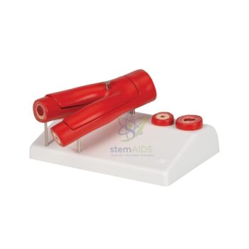
Model, T. S. Monocot Leaf
Histological Tissue Mapping: Features precision-molded representations of bulliform cells, undifferentiated mesophyll, and bundle sheath cells to illustrate the specialized water-storage and photosynthetic mechanics of monocotyledonous species.
Reinforced Structural Relief: The transverse section is mounted on a chemical-resistant polymer baseboard, utilizing high-contrast, medical-grade pigments to differentiate xylem and phloem vessels for intensive tactile examination and high-frequency handling.
₹460.00
Quick Answer: The model, T.S. monocot leaf, is an enlarged transverse section of a monocotyledonous leaf such as maize or wheat. A monocot leaf model shows the isobilateral structure, with similar upper and lower sides, the undifferentiated mesophyll and the row of parallel vascular bundles.
Isobilateral Structure
Grass-like monocot leaves often grow upright, so both surfaces receive similar light. The result is an isobilateral leaf: stomata occur in both the upper and the lower epidermis, and the mesophyll is not divided into palisade and spongy layers but consists of fairly uniform cells with chloroplasts and small air spaces.
Some features appear only in monocots. Groups of large, thin-walled, colourless bulliform cells in the upper epidermis lose water in dry weather, making the leaf roll inwards and reducing water loss. The guard cells of grasses are dumbbell-shaped. Because the veins run parallel, a transverse section cuts many vascular bundles of different sizes in a row, each surrounded by a bundle sheath and often with patches of sclerenchyma above and below.
In C4 grasses such as maize, the bundle sheath cells are large and contain chloroplasts, and the mesophyll forms a ring around them. This Kranz anatomy, from the German word for wreath, is a common extension topic, though whether this model depicts it is not stated. The T.S. dicot leaf model gives the contrast with a dorsiventral leaf. Format and size can be checked with LabEquip.
Care & Handling
- Keep the monocot leaf model upright or flat as designed and support it from below when carrying it.
- Remove dust with a soft brush; do not use wet cloths on the painted section.
- Store it covered, away from heat and light.
Specifications
| Model of | Transverse section (T.S.) of a monocot leaf |
| Leaf type | Isobilateral |
| Tissues shown | Epidermis on both sides with stomata, bulliform cells, mesophyll, parallel vascular bundles, bundle sheath, sclerenchyma |
| Extension topic | Kranz anatomy of C4 grasses |
| Species, format and size | Confirm at enquiry |
What It Teaches
- Comparing isobilateral monocot and dorsiventral dicot leaves
- How bulliform cells let grass leaves roll up in drought
- Introducing C4 photosynthesis and Kranz anatomy in senior biology
- Spotting tests alongside prepared sections of maize or wheat leaf
Why Choose LabEquip
Botany departments buy transverse leaf sections so students can compare the two classes of flowering plants side by side. LabEquip offers this model within its biology lab products, and a monocot leaf chart makes a companion for the wall. Use the LabEquip contact page for orders.
Frequently Asked Questions
What does isobilateral mean?
It means both surfaces of the leaf are alike. In an isobilateral monocot leaf, stomata are present in both epidermal layers and the mesophyll is not divided into palisade and spongy tissue.
What are bulliform cells?
Bulliform cells are large, empty-looking cells in the upper epidermis of grass leaves. When they lose water they shrink, making the leaf roll or fold, which reduces the exposed surface and water loss in dry conditions.
Why are there so many vascular bundles in a monocot leaf section?
Monocot leaves have parallel venation, so the veins run side by side along the length of the leaf. A transverse cut crosses all of them, giving a row of large and small bundles.
What is Kranz anatomy?
It is the wreath-like arrangement in C4 plants such as maize and sugarcane, where large bundle sheath cells with chloroplasts are ringed by mesophyll cells. It concentrates carbon dioxide around the enzyme RuBisCO and improves photosynthesis in hot conditions.
What shape are the guard cells in grasses?
They are dumbbell-shaped, narrow in the middle with swollen ends, and flanked by subsidiary cells. Dicot guard cells, by contrast, are bean- or kidney-shaped.
Which leaves can students section to compare with the model?
Maize, wheat, sugarcane or any lawn grass give clear monocot sections. Thin transverse slices cut with a razor, stained with safranin and mounted in water show the main tissues under a school microscope.
Last Updated: September 2026
You must be logged in to post a review.
Bestsellers




Reviews
There are no reviews yet.