


In Stock
Model of a Cell in Mitosis
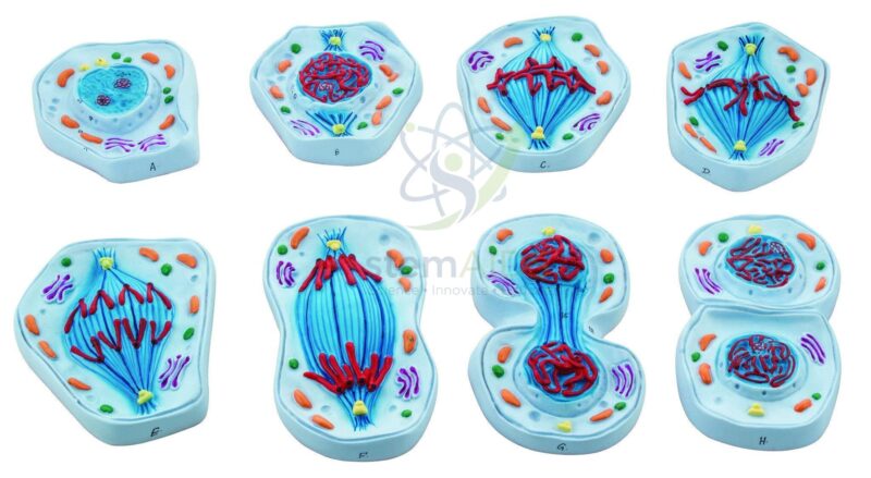
Modèle de cellule en mitose Features eight discrete, high-relief models representing sequential mitotic phases, each with color-coded chromatin and centrosome structures for detailed microscopic-level observation and tactile analysis.
Reinforced Laboratory-Grade Polymer Constructed from high-impact, non-toxic PVC with UV-resistant pigments to ensure long-term dimensional stability and color retention under high-frequency institutional handling and repetitive sterilization cycles.
₹11,250.00
Quick Answer: The Model of a Cell in Mitosis is a mitosis model that shows an animal cell at successive stages of division, from interphase through prophase, metaphase, anaphase and telophase to the two daughter cells formed by cytokinesis. Biology teachers use it to show how copied chromosomes are shared equally between new cells.
Reading the Stages on the Mitosis Model
The product photo shows the model as a series of eight separate cell pieces laid out in order, each with coloured chromosomes, spindle fibres and organelles. The cells have no cell wall, so they represent animal cells. The title names a single cell, so confirm the number of pieces supplied; the stages described here are the ones such a series is designed to show.
In interphase the chromosomes are not visible as separate threads, because the DNA is spread through the nucleus while it is copied. In prophase each chromosome condenses into two sister chromatids joined at a centromere, and the centrosomes move apart as the spindle forms. Once the nuclear envelope has broken down, the chromosomes line up across the middle of the cell at metaphase. At anaphase the sister chromatids are pulled to opposite poles, and at telophase new nuclear envelopes form around each set while a cleavage furrow begins to pinch the cytoplasm in two.
Laying the pieces side by side makes testing easy: shuffle them and ask for the correct order, or point to one piece and ask what has just happened and what comes next. For comparing mitosis with the reduction division that makes gametes, the animal meiosis model follows the same style of teaching.
Classroom Uses
- Sequencing exercises in which students arrange the stages of mitosis in order
- Explaining why each daughter cell receives an identical set of chromosomes
- Contrasting cytokinesis in animal cells with the cell plate formed in plant cells
- Revision before onion root tip practicals, linking model stages to what is seen under the microscope
Care & Handling
- Keep the mitosis model pieces in a divided tray or box in stage order, so a missing piece is noticed at once.
- Handle the pieces by their edges; the raised chromosomes and spindle fibres are the parts most likely to snap.
- Wipe with a soft, dry or barely damp cloth, never with solvents that lift the colours used to tell chromosomes apart.
Specifications
| Model of | Animal cell at successive stages of mitosis |
| Pieces in the product photo | Eight cells, from interphase to two daughter cells |
| Features | Chromosomes, spindle fibres, centrosomes, nucleus and cytoplasmic organelles |
| Cell type | Animal cell: no cell wall, divides by a cleavage furrow |
| Number of pieces, size and material | Confirm at enquiry |
Why Choose LabEquip
Secondary schools and pre-university biology departments buy stage-by-stage mitosis models because students can handle the sequence rather than memorise it from a page. The set is part of LabEquip’s biology lab products; ask about the piece count and size through the contact page.
Frequently Asked Questions
What are the stages of mitosis in order?
Prophase, metaphase, anaphase and telophase, usually followed by cytokinesis. Interphase, when DNA is copied, comes before mitosis and is not itself a stage of it. Some courses also name prometaphase, between prophase and metaphase.
What is the difference between a chromosome and a chromatid?
After DNA is copied, each chromosome consists of two identical sister chromatids held together at the centromere. At anaphase the chromatids separate, and from then on each one is counted as a chromosome in its own right.
How does mitosis differ from meiosis?
Mitosis produces two genetically identical cells with the same chromosome number as the parent, for growth and repair. Meiosis involves two divisions and produces four cells with half the chromosome number, used to make gametes.
Why does this model show an animal cell rather than a plant cell?
The cells have no rigid wall and divide by a cleavage furrow that pinches the cell in two. A dividing plant cell instead builds a cell plate across the middle, which becomes the new wall between the daughter cells.
Which practical goes well with this model?
The onion root tip squash, in which the growing tip of a root is softened, stained and pressed flat on a slide. Students find cells at different stages under the microscope and match them to the model pieces.
What are spindle fibres made of?
They are made of microtubules, hollow protein tubes built from tubulin. They grow out from the centrosomes, attach to the chromosomes at their centromeres and shorten to pull the chromatids apart at anaphase.
Last Updated: September 2026
You must be logged in to post a review.
Bestsellers




Reviews
There are no reviews yet.