


In Stock
Human Hand Model
$28.88
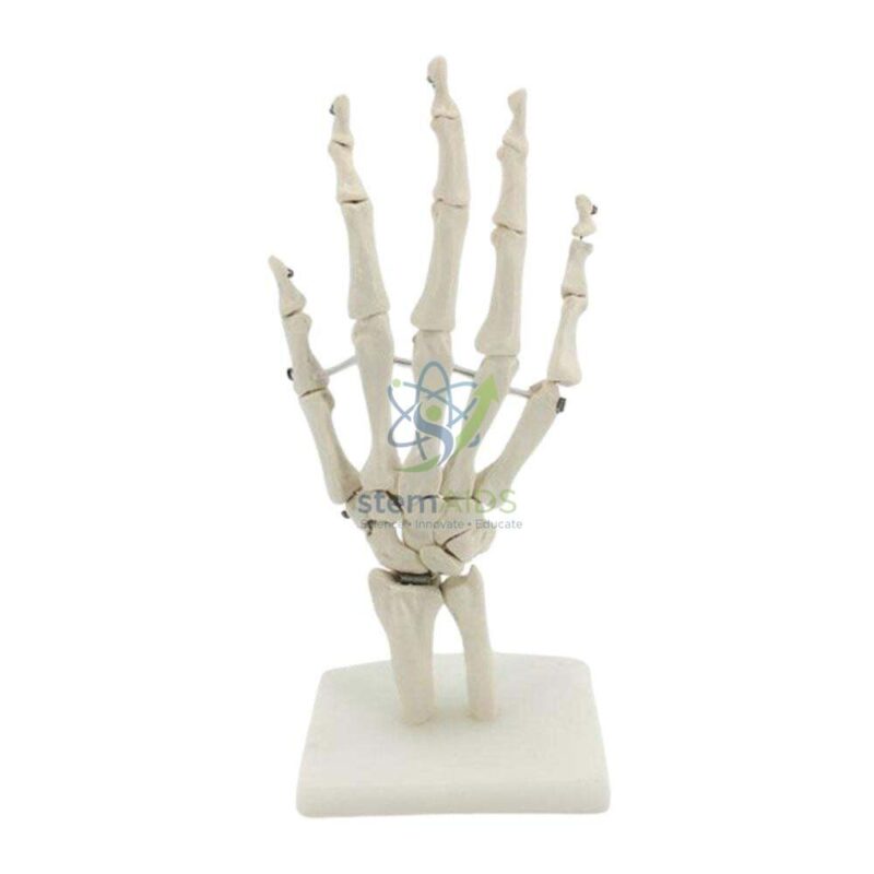
Quick Answer: The Human Hand Model is an anatomy teaching model of the skeleton of the hand. The listing photograph shows the bones of the wrist, palm and fingers articulated on a stand, used to teach the carpals, metacarpals and phalanges and the joints that make the hand so mobile.
Reading the Bones of the Hand
Laid out on a stand, the skeleton of the hand divides into three groups that students can count off in order. At the wrist sit eight small, irregular carpal bones packed in two rows. Five long metacarpals form the palm, one leading to each digit. The fingers have three phalanges each, proximal, middle and distal, while the thumb has only two, giving fourteen phalanges and 27 bones in all.
A popular memory aid for the carpals, starting on the thumb side of the row nearest the forearm and returning along the row nearest the fingers, is ‘So Long To Pinky, Here Comes The Thumb’: scaphoid, lunate, triquetrum, pisiform, hamate, capitate, trapezoid, trapezium. The model makes the aid concrete, and it shows why the scaphoid, bridging both rows on the thumb side, is the carpal most often broken in a fall on an outstretched hand.
The hand also shows several joint types in a small space. The joints between phalanges are hinges that only bend and straighten; the knuckles allow bending plus side-to-side spreading; and the thumb’s base is a saddle joint that lets it swing across the palm to meet each fingertip. Whether this model includes the ends of the forearm bones, how the bones are joined and its scale are not stated in the title. For soft tissue, the hand joint model with ligaments adds the ligaments that bind these bones together.
Specifications
| Model of | Skeleton of the human hand and wrist |
| Format | Articulated bones on a stand, per the listing photograph |
| Bones shown | Carpals, metacarpals and phalanges |
| Topics | Bone names, joint types, common hand fractures |
| Scale, material and forearm bones | Confirm at enquiry |
Applications
- Naming and counting the bones of the hand in secondary biology
- Comparing hinge, condyloid and saddle joints in one structure
- Explaining common injuries such as scaphoid and fifth metacarpal fractures
- Helping nursing, physiotherapy and first-aid students read hand X-ray images
Care & Handling
- Carry the model by its stand, not by the fingers, which are thin and can snap at the joints.
- Dust with a soft brush; avoid bending wired joints further than they move naturally.
- Store it covered and away from sunlight and heat, which can yellow or warp plastic bones.
Why Choose LabEquip
Anatomy teachers often order a hand and a foot model together to compare the two limbs. LabEquip lists this model in Biology Lab Products with the matching Human Foot Model. Use the LabEquip contact page to confirm scale and how the bones are mounted.
Frequently Asked Questions
How many bones are in the human hand?
There are 27: eight carpals in the wrist, five metacarpals in the palm and fourteen phalanges in the digits. Some sources add small sesamoid bones, which vary between people.
Why does the thumb have only two phalanges?
The thumb has proximal and distal phalanges but no middle one. Its long metacarpal and the saddle joint at its base make up for this, letting it rotate across the palm to grip.
Which wrist bone is most often fractured?
The scaphoid, on the thumb side of the wrist, is the carpal most often broken, usually in a fall on an outstretched hand. The model shows how it links the two rows of carpals, which is why it takes the force.
What joint types can students identify on the model?
Hinge joints between the phalanges, condyloid joints at the knuckles that also allow spreading, and the saddle joint at the base of the thumb. Movements between carpals are small gliding movements.
How does this model differ from a hand model with ligaments?
This model shows the bones and their arrangement. A ligament model adds the bands of tissue that hold the joints together, which helps when teaching sprains and carpal tunnel anatomy.
How can the model help with reading hand X-rays?
Students hold the model in the same position as the X-ray and match each shadow to a bone. It helps them recognise normal outlines before they look for fractures or dislocations.
Last Updated: September 2026
You must be logged in to post a review.
Bestsellers




Reviews
There are no reviews yet.