
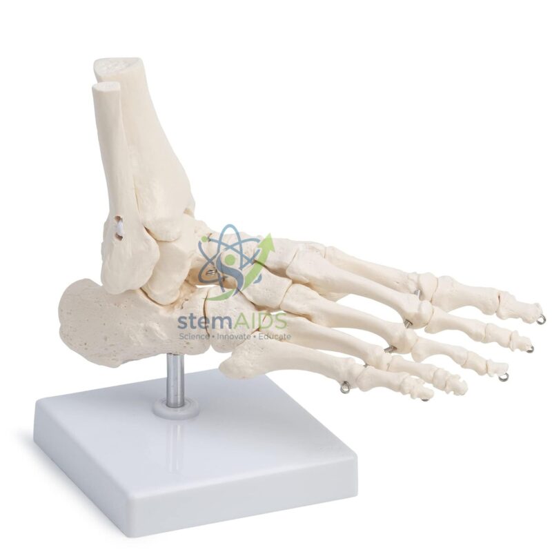
Quick Answer: The Human Foot Model is an anatomy teaching model of the skeleton of the foot. The listing photograph shows the tarsal bones, metatarsals and toe bones articulated on a stand, used to teach how the foot bears weight, how the ankle joint works and where common injuries occur.
A Skeleton Built for Standing
The foot does two jobs the hand never has to: it carries the whole body’s weight and it acts as a springy lever for walking and running. Its skeleton reflects this. Seven tarsal bones, much larger than the carpals of the wrist, form the heel and the back of the foot. The talus sits on top and fits into the socket formed by the lower ends of the tibia and fibula at the ankle; beneath it the calcaneus forms the heel and takes the first impact of each step. In front lie the navicular, the cuboid and three cuneiforms, then five metatarsals and fourteen phalanges.
The model shows how these bones are arranged in an arch rather than lying flat. Weight passes down through the talus and spreads backwards to the heel and forwards to the heads of the metatarsals, which is why these points bear pressure while the middle of the inner foot stays off the ground. The big toe, unlike the thumb, is aligned with the other toes, a design for push-off rather than grasping.
It shows bone only; ligaments, tendons and the plantar fascia are not represented. For comparing normal, flat and high-arched feet, LabEquip lists a separate human foot set model.
Teaching Uses
- Naming the tarsals and tracing the path of body weight through the foot
- Comparing the skeleton of the foot with that of the hand
- Explaining ankle fractures, metatarsal stress fractures and bunions
- Physiotherapy, sports-science and podiatry introductions
Specifications
| Model of | Skeleton of the human foot |
| Format | Articulated bones on a stand, per the listing photograph |
| Bones shown | Tarsals, metatarsals and phalanges |
| Topics | Ankle joint, arches, weight bearing, common fractures |
Care & Handling
- Lift the model by its base; the small toe bones are fragile at their joints.
- Clean with a soft brush or a slightly damp cloth, never with solvents.
- Keep the model in a cupboard or under a cover so the fine joints do not collect dust.
Why Choose LabEquip
Schools, nursing colleges and sports-science courses often buy a foot model with a hand model for limb comparison. LabEquip lists this model in Biology Lab Products beside the Human Hand Model. Ask about scale and mounting through the LabEquip contact page.
Related Products
- Human Foot Set Model: A foot set model for comparing normal, flat and high-arched feet and the three arches.
- Human Hand Model: The matching articulated hand skeleton on a stand, for comparing hand and foot bones.
- Foot Bone Anatomy Model: Another foot bone model for the lower-limb skeleton, a second option for group work.
- Human Leg Bone Model With Blood Vessels: Leg bones with blood vessels, including the dorsalis pedis artery on top of the foot.
Frequently Asked Questions
How many bones are in the human foot?
Each foot has 26 bones: 7 tarsals, 5 metatarsals and 14 phalanges. This foot model shows them articulated on a stand, so students can count and name them in their natural arrangement.
Which bones form the ankle joint?
The ankle joint is formed by the talus fitting into a socket made by the lower ends of the tibia and fibula. It works mainly as a hinge for pointing the foot up and down; turning the sole inwards and outwards happens at joints below the talus.
What are the names of the tarsal bones?
The seven tarsals are the talus, calcaneus (heel bone), navicular, cuboid and the medial, intermediate and lateral cuneiforms. The talus and calcaneus form the back of the foot and take the body’s weight first in each step.
How is the foot skeleton different from the hand skeleton?
The foot has larger, stronger tarsal bones built to carry weight, while the wrist’s carpals are small and allow fine movement. The big toe lines up with the other toes and cannot be opposed like the thumb, so the foot is built for support and push-off rather than grip.
Where do stress fractures in the foot usually happen?
They most often affect the shafts of the second and third metatarsals, sometimes called march fractures, after repeated impact from running or long marches. The navicular and the base of the fifth metatarsal are other sites that the model helps students locate.
How is this foot model different from a foot set model?
This model is a single articulated foot skeleton for learning bone names, joints and injury sites. A foot set is used to compare foot shapes, such as a normal arch, a flat foot and a high arch.
Last Updated: November 2026




Reviews
There are no reviews yet.