
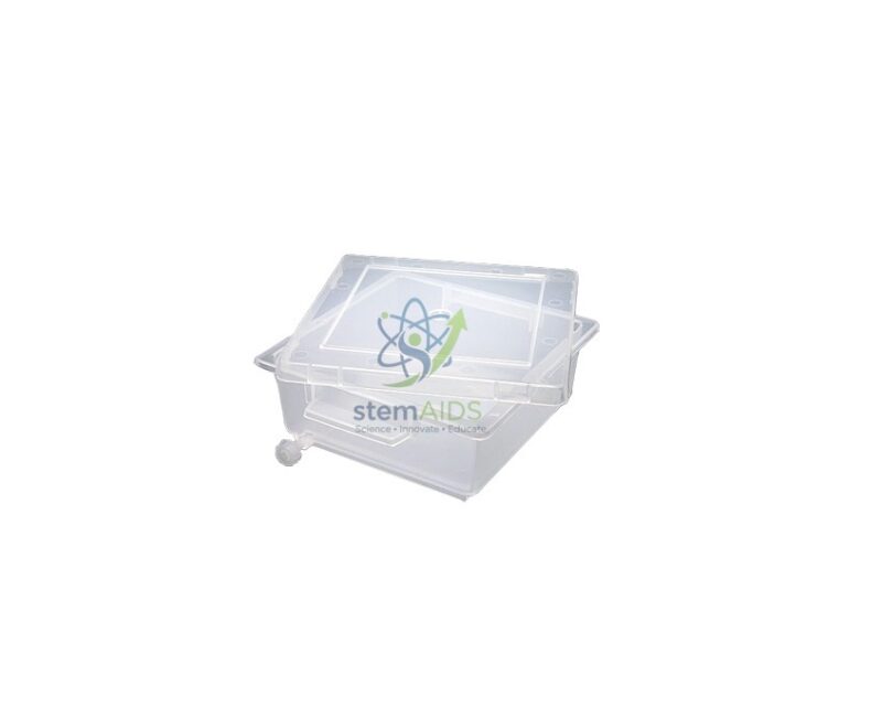
Quick Answer: A gel staining tray is a shallow, lidded plastic container in which an electrophoresis gel is soaked in stain and then destained. The listing photograph shows a clear tray with a clear lid, used after agarose gel electrophoresis to make DNA bands visible.
Where the Tray Fits in an Electrophoresis Practical
An agarose gel straight out of the electrophoresis tank shows nothing but the loading dye. DNA fragments are colourless, so after the run the gel is slid into a staining tray, covered with stain and left to soak. The stain binds to the DNA; the gel is then moved into water, often in the same tray after the stain is poured off, until the background clears and the bands stand out. A lid keeps dust out during long soaks, slows evaporation and lets the tray be carried or stacked without spills.
Schools often use methylene blue or other stains that can be viewed under ordinary light, because they avoid ultraviolet viewing. College and research labs may use fluorescent DNA stains, which are viewed on a transilluminator, or Coomassie-type stains for protein gels. Before using solvent-based stains or destain solutions, check that the plastic of this tray tolerates them; the title does not name the plastic.
A flat, clear tray also helps the handling itself. Agarose gels of low concentration tear easily, and a tray a little larger than the gel lets it be tilted, drained and photographed without lifting it more than necessary. Tray dimensions are not stated, so match them to the size of gel your casting trays produce.
Applications
- Staining agarose gels from restriction-digest or DNA-fingerprinting simulation practicals
- Destaining gels in several changes of water until bands are clearly visible
- Carrying a stained gel from the bench to a light box or camera stand
- Keeping gels covered overnight between two practical sessions
Specifications
| Item | Tray with lid for staining and destaining electrophoresis gels |
| Construction | Clear plastic tray and lid, as shown in the listing photograph |
| Typical gels | Agarose DNA gels; protein gels where the stain suits the plastic |
| Use steps | Stain, pour off, destain in water, view or photograph |
Care & Handling
- Wear gloves when handling stained gels and stain solutions; methylene blue and other dyes mark skin and clothing.
- Keep separate, labelled trays for different stains so traces of one do not tint the next gel.
- Rinse the tray and lid soon after use with water and a mild detergent, and dry them without abrasive pads that scratch the clear plastic.
Why Choose LabEquip
Staining trays are usually ordered with electrophoresis kits for biotechnology practicals in senior school and first-year college. LabEquip lists the tray in its Biology Lab Products range beside teaching models such as the DNA PCR principle model. Share your gel size and stain through the LabEquip contact page so the tray can be matched.
Related Products
- Staining Box: A staining box for batch immersion of microscope slide racks, the slide counterpart of the gel tray.
- Pipette, Pasteur, Disposable: Disposable Pasteur pipettes for adding and drawing off small volumes of stain and water.
- Molecular Model of DNA PCR Principle: A model of the polymerase chain reaction, whose products are often checked on an agarose gel.
- Gloves: Protective gloves for handling stained gels and keeping stain off the hands.
Frequently Asked Questions
What is a gel staining tray used for?
After electrophoresis the gel is slid into the tray, covered with stain and left to soak; then the stain is poured off and the gel destained in water until the bands stand out. The listing photo shows a clear tray with a clear lid.
Which stains can be used in a gel staining tray?
Schools often use methylene blue or similar stains that show DNA bands under ordinary light. Research labs use fluorescent DNA stains viewed under blue or UV light, and Coomassie blue for protein gels. Make sure the stain and any destaining solvent suit the tray’s plastic.
How long should an agarose gel be stained and destained?
Times depend on the stain, its strength and the gel thickness, so follow the stain’s own method. With visible-light DNA stains, staining is usually the short step and destaining in several changes of water takes longer, until the background is pale and the bands are sharp.
How do you move an agarose gel into a staining tray without breaking it?
Keep the gel on its casting tray and slide it gently off into the liquid, or support it underneath with a flat spatula or a gloved palm. Never lift it by one edge: thin gels with a low agarose content tear easily.
Can a gel be viewed on a UV transilluminator in the staining tray?
Most clear plastics absorb UV light, which dims the image, so the gel is usually moved onto the transilluminator glass. For visible-light stains such as methylene blue, the tray can be set on a white light box instead.
Why does a gel staining tray have a lid?
The lid keeps dust out of the stain, slows evaporation during long soaks and lets the tray be carried or stacked without spilling. It also cuts down splashes and vapour from the staining solution.
Last Updated: November 2026




Reviews
There are no reviews yet.