

In Stock
Gel Staining Tray
₹335.00
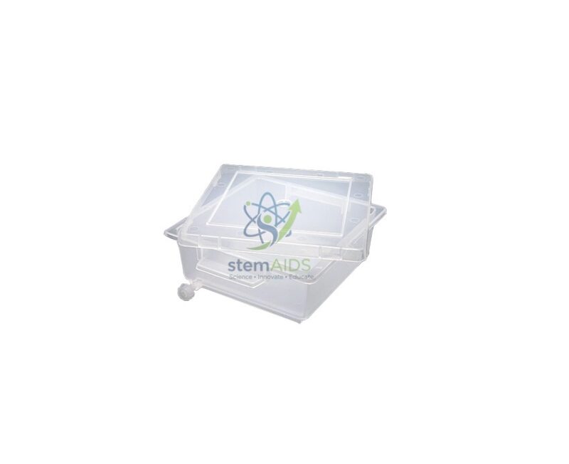
Quick Answer: A gel staining tray is a shallow, lidded plastic container in which an electrophoresis gel is soaked in stain and then destained. The listing photograph shows a clear tray with a clear lid, used after agarose gel electrophoresis to make DNA bands visible.
Where the Tray Fits in an Electrophoresis Practical
An agarose gel straight out of the electrophoresis tank shows nothing but the loading dye. DNA fragments are colourless, so after the run the gel is slid into a staining tray, covered with stain and left to soak. The stain binds to the DNA; the gel is then moved into water, often in the same tray after the stain is poured off, until the background clears and the bands stand out. A lid keeps dust out during long soaks, slows evaporation and lets the tray be carried or stacked without spills.
Schools often use methylene blue or other stains that can be viewed under ordinary light, because they avoid ultraviolet viewing. College and research labs may use fluorescent DNA stains, which are viewed on a transilluminator, or Coomassie-type stains for protein gels. Before using solvent-based stains or destain solutions, check that the plastic of this tray tolerates them; the title does not name the plastic.
A flat, clear tray also helps the handling itself. Agarose gels of low concentration tear easily, and a tray a little larger than the gel lets it be tilted, drained and photographed without lifting it more than necessary. Tray dimensions are not stated, so match them to the size of gel your casting trays produce.
Applications
- Staining agarose gels from restriction-digest or DNA-fingerprinting simulation practicals
- Destaining gels in several changes of water until bands are clearly visible
- Carrying a stained gel from the bench to a light box or camera stand
- Keeping gels covered overnight between two practical sessions
Specifications
| Item | Tray with lid for staining and destaining electrophoresis gels |
| Construction | Clear plastic tray and lid, as shown in the listing photograph |
| Typical gels | Agarose DNA gels; protein gels where the stain suits the plastic |
| Use steps | Stain, pour off, destain in water, view or photograph |
| Dimensions and plastic type | Confirm at enquiry |
Care & Handling
- Wear gloves when handling stained gels and stain solutions; methylene blue and other dyes mark skin and clothing.
- Keep separate, labelled trays for different stains so traces of one do not tint the next gel.
- Rinse the tray and lid soon after use with water and a mild detergent, and dry them without abrasive pads that scratch the clear plastic.
Why Choose LabEquip
Staining trays are usually ordered with electrophoresis kits for biotechnology practicals in senior school and first-year college. LabEquip lists the tray in its Biology Lab Products range beside teaching models such as the DNA PCR principle model. Share your gel size and stain through the LabEquip contact page so the tray can be matched.
Frequently Asked Questions
Which stains can be used in a gel staining tray?
Methylene blue is common in schools because the bands are seen in ordinary light. Fluorescent DNA stains and protein stains such as Coomassie are used in colleges. Check that the tray plastic suits any stain or destain that contains alcohol, acid or other solvents.
How long does a gel need to stay in the stain?
It depends on the stain, the gel thickness and the agarose concentration. Many teaching protocols with methylene blue stain for around half an hour and then destain in several changes of water, sometimes overnight. Follow the protocol supplied with the stain.
Why does the tray need a lid?
A lid keeps dust and debris out during long soaks, slows evaporation of the stain, reduces the chance of spills when the tray is moved, and keeps light-sensitive stains shaded. It also lets trays be stacked while gels wait between lessons.
How do I move an agarose gel into the tray without breaking it?
Slide the gel off its casting tray directly into the staining tray, or support it on a gloved flat hand. Avoid lifting it by one edge. Low-percentage gels are especially fragile and are safer moved on the casting tray itself.
Can the tray be placed on a UV transilluminator?
Many plastics absorb ultraviolet light and would dim the bands. The usual practice is to transfer the gel from the tray onto the transilluminator surface for viewing. Always use UV face protection when a transilluminator is on.
Can one tray be used for both DNA and protein gels?
It can if it is cleaned well between uses and the plastic suits both stains, but most labs keep dedicated trays for each stain. This avoids carry-over of dye and makes it clear which waste stream the used solution belongs to.
Last Updated: September 2026
You must be logged in to post a review.
Bestsellers




Reviews
There are no reviews yet.