

Quick Answer: The fetus with viscera and placenta is a model of a fetus with its internal organs (viscera) exposed, joined by the umbilical cord to a placenta. It is used in nursing, midwifery and medical teaching to show fetal anatomy and, above all, how fetal circulation differs from circulation after birth.
Fetal Circulation, Organ by Organ
The value of showing the viscera is that the fetal shunts can be traced. Oxygen-rich blood leaves the placenta in the single umbilical vein and reaches the liver, where much of it bypasses the liver tissue through the ductus venosus into the inferior vena cava. In the heart, blood passes from the right atrium to the left through the foramen ovale, and much of the blood pumped into the pulmonary trunk crosses to the aorta through the ductus arteriosus, because the fluid-filled lungs need little flow. Two umbilical arteries return blood to the placenta.
At birth these shunts close. The first breaths expand the lungs and lower resistance in the pulmonary vessels, pressure in the left atrium rises and pushes the flap of the foramen ovale shut, and the ductus arteriosus constricts over the following hours to days. The ductus venosus and the umbilical vessels later become ligaments. A model showing the heart, liver, lungs and cord together is a clear way to teach this change.
Viscera models also show fetal proportions: the liver is very large and fills much of the abdomen, and the head is big relative to the body. The gestational age represented, the scale and whether organs can be removed are not stated in the title; the related human placenta model shows the placental surfaces in more detail.
Care & Handling
- Support both the fetus and the placenta when lifting the model so the cord connection is not strained.
- Clean the exposed organs with a soft dry brush; damp cloths can lift paint from fine vessel detail.
- Store it flat in its box with any removable organs replaced.
Specifications
| Model of | Fetus with internal organs exposed, joined to a placenta |
| Connection | Umbilical cord with its vessels, as usually modelled |
| Teaching focus | Fetal circulation, fetal organ proportions, placenta |
| Users | Midwifery, nursing, neonatal and medical students |
Applications
- Teaching fetal circulation and the changes at birth in midwifery and neonatal nursing
- Explaining congenital heart conditions linked to the shunts, such as patent ductus arteriosus
- Anatomy and embryology lectures on fetal organ proportions
- Antenatal education on how the baby is nourished before birth
Why Choose LabEquip
Midwifery schools and neonatal nursing programmes buy this kind of model because fetal circulation is difficult to grasp from a flat diagram alone. The model is part of LabEquip’s biology lab products; questions about the gestational age shown can go through the contact page.
Related Products
- Human Placenta: A detailed placenta model with fetal and maternal surfaces and the umbilical cord vessels.
- Embryo Model: An enlarged early embryo, the stage before the fetal period shown on this model.
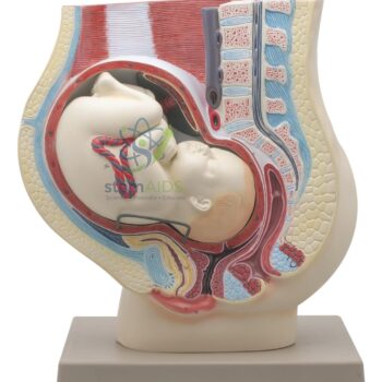
- Pregnant Woman’s Belly: A section through a woman in late pregnancy, with the fetus lying head down in the uterus.
- Human Skull Model, Fetal Child: A fetal skull with fontanelles and sutures, used in midwifery teaching with the fetal anatomy shown here.
Frequently Asked Questions
What are the three shunts in fetal circulation?
The ductus venosus carries much of the blood from the umbilical vein past the liver into the inferior vena cava. The foramen ovale lets blood cross from the right atrium to the left. The ductus arteriosus sends blood from the pulmonary trunk into the aorta, bypassing the fluid-filled lungs.
Why does the umbilical vein carry oxygen-rich blood?
Vessels are named by the direction of flow relative to the heart, not by oxygen content. The umbilical vein carries blood from the placenta, where it has picked up oxygen, towards the fetal heart; the two umbilical arteries carry blood from the fetus back to the placenta.
What happens to the foramen ovale and ductus arteriosus after birth?
With the first breaths, resistance in the lungs falls and pressure in the left atrium rises, pushing the flap of the foramen ovale shut; it later seals as the fossa ovalis. The ductus arteriosus narrows within days and becomes the ligamentum arteriosum.
Why is the fetal liver so large?
For much of pregnancy the fetal liver is a major site of blood cell production, and it receives blood straight from the umbilical vein, so it takes up a large share of the abdomen. The exposed viscera of this model make that proportion easy to see.
What does the placenta do for the fetus?
The placenta is where oxygen and nutrients pass from the mother’s blood to the fetus and carbon dioxide and wastes pass back, without the two blood supplies mixing. It also makes hormones that maintain the pregnancy. On this model it is joined to the fetus by the umbilical cord.
Who uses a fetus with viscera and placenta model?
Midwifery, nursing, neonatal and medical students use it, mainly to learn fetal circulation and fetal organ proportions. It also helps explain to parents why some newborns need their heart checked after birth.
Last Updated: November 2026








Reviews
There are no reviews yet.