


In Stock
Detachable Eye
Œil démontable features a precision-engineered detachable sclera, cornea, and iris, allowing for a three-dimensional examination of internal vitreous humor dynamics and optic nerve positioning.
Multi-Chambered Optical Fidelity incorporates an interchangeable crystalline lens and a high-resolution representation of the vascularized retina, calibrated to a 5x life-size scale for enhanced tactile analysis in institutional laboratory settings.
₹8,560.00
Quick Answer: The Detachable Eye is a detachable eye model: an enlarged human eyeball on a stand that comes apart so the inner layers, lens and chambers can be examined. It is used in biology, optometry foundation and nursing lessons on the structure of the eye and how it focuses light.
Taking the Detachable Eye Model Apart
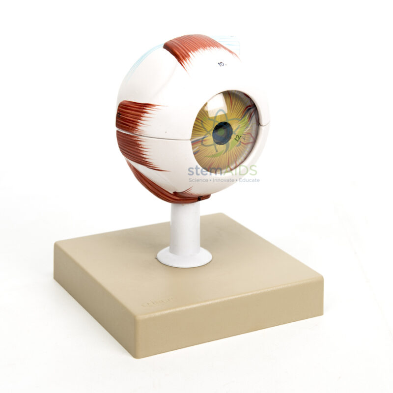
The product photo shows the eyeball on a white stem above a square base, with the extraocular muscles attached to the outside of the white sclera and the cornea and iris at the front. A seam around the middle of the globe marks where the model separates. Dissectible eye models of this kind usually open into upper and lower halves of the sclera, revealing the choroid and retina lining the back of the eye, with the lens and vitreous body removable from inside. The listing gives neither the number of parts nor the magnification, so confirm both at enquiry.
Opening the model in order follows the path of light. Light first meets the clear, curved cornea, responsible for most of the eye’s focusing power, then crosses the aqueous humour and the pupil, whose size the iris controls. The lens fine-tunes focus by changing shape: the ciliary muscle contracts to let it thicken for near objects, a process called accommodation. Light then crosses the vitreous body and forms an image on the retina, where rods and cones turn it into nerve signals that leave through the optic nerve.
The muscles on the outside explain how both eyes move together: four straight (rectus) muscles and two oblique muscles turn the eye up, down, inward, outward and around its axis. For a model that adds the eyelids and surrounding structures, see the human eye model with eyelids.
Lessons on Vision
- Tracing the path of light through the eye in secondary biology
- Explaining accommodation and why near vision becomes harder with age
- Relating short and long sight to where the image falls relative to the retina
- Locating the blind spot at the optic disc
- Nursing and optometry foundation lessons on eye structure and eye movements
Specifications
| Model of | Enlarged human eyeball that separates into parts |
| Mounting | On a stem and base, as shown in the product photo |
| External features | Sclera, cornea, iris and extraocular muscles |
| Internal structures typically shown | Choroid, retina, lens, vitreous body and optic nerve |
| Number of parts and magnification | Confirm at enquiry |
Care & Handling
- Separate and rejoin the halves of the detachable eye model gently along the seam; forcing them wears the locating pins.
- Keep the lens and other small internal parts in a labelled bag or tray while the model is open.
- Wipe with a soft cloth; the clear cornea scratches easily, so avoid paper towels and abrasive cleaners.
Why Choose LabEquip
Biology teachers and optometry and nursing tutors choose a detachable eye because learners can open it and see layers that a whole-eye model hides. It is part of LabEquip’s biology lab products; for part counts and size, send a query through the contact page.
Frequently Asked Questions
Is the cornea or the lens more important for focusing?
The cornea supplies most of the focusing power, because light bends sharply as it passes from air into its curved surface. The lens adds the adjustable part, changing shape so that both near and distant objects form a sharp image on the retina.
What is accommodation?
Accommodation means adjusting the lens so the eye stays in focus as an object’s distance changes. For close work the ciliary muscle contracts, the zonule fibres holding the lens go slack and the elastic lens rounds up; for distant objects the muscle relaxes and the fibres pull the lens flatter.
Why does the eye have a blind spot?
Where the optic nerve and blood vessels leave the back of the eye, the retina has no photoreceptors, so light falling there is not detected. The brain fills in the gap, which is why it normally goes unnoticed.
How do short sight and long sight relate to the model?
In short sight the eye focuses distant light in front of the retina, often because the eyeball is too long. In long sight the focus falls behind the retina. Concave or convex spectacle lenses move the focus back onto it.
How many muscles move the eye?
Six extraocular muscles move each eye: the superior, inferior, medial and lateral rectus muscles and the superior and inferior oblique muscles. They work together so both eyes point at the same object.
How is this model different from an eye model with eyelids?
This model centres on the eyeball itself and comes apart to show its inner layers. A model with eyelids adds the lids and surrounding structures, which suits lessons on how the eye is protected.
Last Updated: September 2026
You must be logged in to post a review.
Bestsellers




Reviews
There are no reviews yet.