
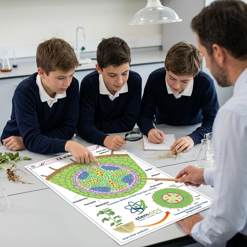
Quick Answer: The Dicot Root Structure Chart is a botany wall chart showing the internal anatomy of a dicotyledonous root in transverse section, usually a gram, bean or sunflower root, with its root-hair layer, cortex, endodermis, pericycle and a small number of radial xylem strands. It supports root anatomy lessons and microscope practicals.
Inside a Young Dicot Root
A dicot root in section looks deceptively simple: a wide cortex around a small, tightly packed stele. The chart typically starts at the epiblema, the single outer layer that bears root hairs, then shows the cortex of rounded parenchyma cells with air spaces between them. The endodermis at the cortex’s inner edge is drawn with Casparian strips, the bands that make water pass through the cells rather than around them.
The stele is where the dicot pattern shows. Inside a one-layered pericycle, xylem and phloem lie on alternate radii, with only two to four xylem strands (diarch to tetrarch) that meet or nearly meet at the centre, so the pith is small or absent. The parenchyma between xylem and phloem, the conjunctive tissue, is often labelled because it matters later.
That later stage is secondary growth. In older dicot roots, cambium arises from the conjunctive tissue and from pericycle cells opposite the protoxylem, joins into a wavy ring and adds secondary xylem and phloem, while a cork layer replaces the cortex. Some charts show this as a second diagram; the panels differ between editions.
Specifications
| Section | Transverse section of a young dicotyledonous root |
| Plants usually drawn | Gram, bean or sunflower root |
| Cortex and boundary | Epiblema, parenchymatous cortex, endodermis with Casparian strips, pericycle |
| Vascular arrangement | Radial bundles, exarch xylem, two to four xylem strands |
| Distinctive label | Conjunctive tissue, where vascular cambium later forms |
| Chart size and finish | Confirm at enquiry |
Care & Handling
- Keep the dicot root chart on the botany wall for the whole anatomy unit rather than rolling it after one lesson, since root, stem and leaf sections are revised together.
- Remove dust with a dry, soft cloth; water marks show badly on printed charts.
- Avoid pinning through the diagram area; if the chart must be pinned, use the margins.
Applications
- Matching the chart’s labels to a stained section of gram or bean root cut by the students
- Teaching the pathway of water uptake, including the role of the Casparian strip
- Explaining how a dicot root starts secondary growth from conjunctive tissue and pericycle
- Revision sessions comparing dicot root, dicot stem and monocot root in one lesson
Why Choose LabEquip
Teachers who stock this chart usually want the complete plant-organ set: the Transverse Section of Monocot Root Chart for comparison and the Dicot Stem Structure Chart to show how xylem position reverses between root and stem. LabEquip supplies these through its Educational Charts range, and a department order can be sent through the contact page.
Frequently Asked Questions
How many xylem strands does a dicot root have?
Usually two to four, described as diarch, triarch or tetrarch, and occasionally up to six. The number varies with the species, so a gram root and a bean root may not match exactly, but it is always fewer than in the polyarch roots of monocots such as maize.
Why is the pith small or missing in a dicot root?
The metaxylem of each strand extends towards the centre, and the strands often meet there, filling the middle of the stele with xylem. In a monocot root the many xylem strands stay near the edge of the stele, leaving room for a large pith.
What does the pericycle do in a dicot root?
It is a layer of cells just inside the endodermis that keeps the ability to divide. Lateral roots grow from it, and during secondary growth it contributes to the vascular cambium and later to the cork cambium, so it is far more active than its single thin layer suggests.
What is conjunctive tissue?
It is the parenchyma that fills the spaces between the xylem and phloem strands in the stele. In dicot roots it matters because the vascular cambium first appears in this tissue, just inside the phloem, when secondary growth begins.
Does the chart apply to every dicot root?
It shows the general plan of a young dicot root, which holds for most dicots at the primary stage. The number of xylem strands and the thickness of the cortex vary between species, so students sectioning a different plant should expect the same tissues in the same order but not identical proportions.
How can students make a dicot root section?
Germinate gram or bean seeds on moist cotton for a few days, then cut thin transverse slices of the young root, supporting it in a slit piece of carrot. Stain with safranin, mount in glycerine and look for the small, star-shaped xylem at the centre.
Last Updated: September 2026



