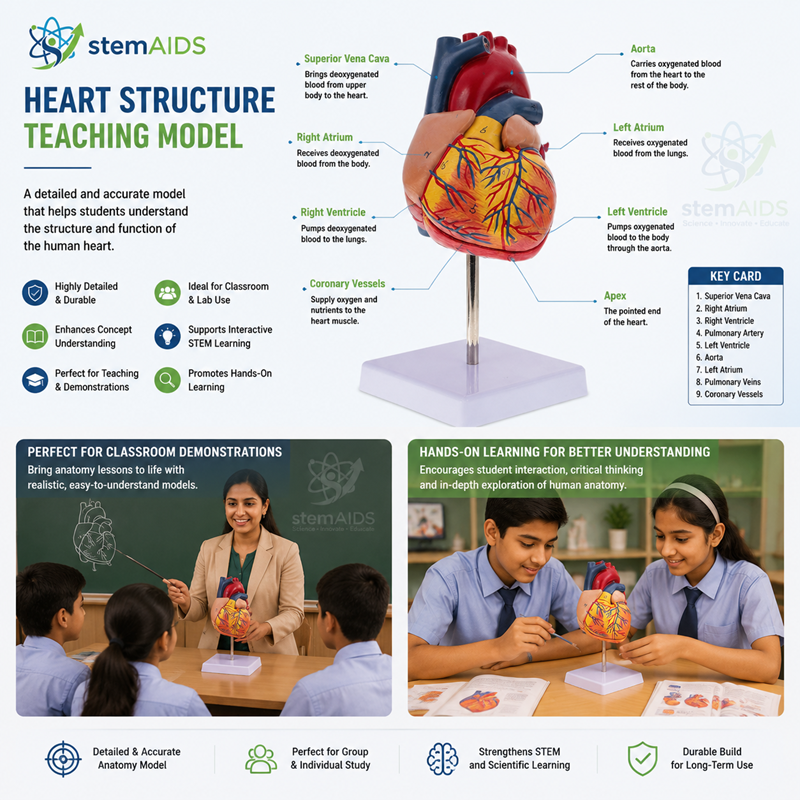
Quick Answer: The Heart Structure Teaching Model is a three-dimensional human heart model for biology and anatomy lessons. It shows how the chambers, valves and major blood vessels of the heart are arranged, so students can follow the path blood takes through them.
Chambers, Valves and Vessels
The human heart has four chambers: two upper atria that receive blood and two lower ventricles that pump it out. A wall of muscle, the septum, separates the right side, which handles deoxygenated blood, from the left side, which handles oxygenated blood. With the Heart Structure Teaching Model on the bench, students can follow which vessel joins which chamber instead of memorising a flat diagram.
The usual route taught is: venae cavae into the right atrium, through the tricuspid valve into the right ventricle, out through the pulmonary artery to the lungs, back through the pulmonary veins to the left atrium, through the bicuspid (mitral) valve into the left ventricle and out through the aorta to the body. The semilunar valves at the base of the pulmonary artery and aorta stop blood flowing back into the ventricles.
Two points regularly come up in exams. The left ventricle has a much thicker muscular wall than the right because it pumps blood around the whole body rather than only to the lungs, and the pulmonary artery is the one artery that carries deoxygenated blood. The coronary arteries on the heart’s surface, which supply the heart muscle itself, open a discussion of heart attacks and healthy lifestyles.
Specifications
| Item | Heart structure teaching model, 3D |
| Features represented | Chambers, valves and major blood vessels of the heart |
| Lessons | Circulatory system, double circulation, cardiac cycle |
| Level | School and college biology and anatomy |
Applications
- Tracing the double circulation through the heart and lungs
- Explaining valve action and the ‘lub-dub’ heart sounds
- Comparing the wall thickness of atria and ventricles
- Pulse-rate practicals before and after exercise
Care & Handling
- Handle the heart model by its main body, not by the projecting vessels.
- Wipe it with a slightly damp cloth after a class has used it and allow it to dry fully.
- Keep it on a shelf at room temperature, out of direct sunlight.
Why Choose LabEquip
Circulation and breathing are usually taught together, so schools often order this heart model with the Human Pulmonary System Model to show where the pulmonary circuit leads. Each is available in the Anatomy Models section, and a biology department can arrange its order on the contact page in one go.
Related Products
- Jumbo Heart Anatomical Replica: An enlarged jumbo heart for whole-class demonstrations from the front of the room.
- Human Heart, 4 Parts: A heart that separates into four parts, giving separate views of the atria, ventricles and valves.
- Human Circulation Educational Model: Places the heart in the double circulation with its major vessels, the next step after its internal structure.
- Digital Heart Rate Monitor: A digital heart rate monitor for the pulse and exercise practicals that usually follow a heart lesson.
Frequently Asked Questions
What is the path of blood through the heart?
Deoxygenated blood enters the right atrium from the venae cavae, passes the tricuspid valve into the right ventricle and leaves through the pulmonary artery to the lungs. Oxygenated blood returns in the pulmonary veins to the left atrium, passes the mitral valve into the left ventricle and is pumped out through the aorta. Tracing this route chamber by chamber is a standard exam exercise.
Why is the left ventricle wall thicker than the right?
The left ventricle pumps blood through the systemic circulation to every part of the body, which needs much higher pressure than the short trip from the right ventricle to the nearby lungs. Its thicker muscle wall generates that pressure. Keeping pressure low on the right side also protects the delicate capillaries in the lungs.
What happens during the cardiac cycle?
Each heartbeat has three stages. In atrial systole the atria contract and push blood into the ventricles; in ventricular systole the ventricles contract, the valves between atria and ventricles shut and blood is forced into the arteries; in diastole the muscle relaxes and the chambers refill. At a resting rate of about 75 beats a minute, one cycle takes roughly 0.8 seconds.
Why does the pulmonary artery carry deoxygenated blood?
Arteries are defined by the direction of flow, not by oxygen content: they carry blood away from the heart. The pulmonary artery takes blood from the right ventricle to the lungs to pick up oxygen, so it carries deoxygenated blood, while the pulmonary veins bring oxygenated blood back to the left atrium. Examiners ask about this exception often.
What is the septum of the heart?
The septum is the wall of muscle that divides the heart into right and left sides. It keeps deoxygenated blood on the right separate from oxygenated blood on the left, so blood sent to the body carries as much oxygen as possible. A hole in the septum, one of the heart defects that can be present at birth, lets the two mix.
Which simple practicals go well with a heart structure lesson?
Taking the pulse at the wrist and neck, measuring heart rate before and after exercise, and listening to heart sounds with a stethoscope all link directly to the chambers and valves on the model. Where a dissection is possible, students can then find the same structures on a real heart and compare the thickness of the two ventricle walls.
Last Updated: November 2026