

Quick Answer: The human pelvis, female, is a replica of the adult female pelvic girdle, the hip bones, sacrum and coccyx, used to teach obstetric landmarks and the dimensions of the birth canal. Midwifery, nursing and anatomy students use a female pelvis model to understand how the shape of the pelvis guides a baby through labour.
The Female Pelvis in Obstetric Teaching
Midwifery courses treat the pelvis as a canal with three levels: the inlet at the pelvic brim, the curved cavity, and the outlet, framed by the subpubic arch in front, the ischial tuberosities at the sides and the coccyx behind. Each level has a longest and a shortest diameter, and the fetal head turns as it descends so that its own longest dimension meets the widest space available. A model makes this rotation far easier to follow than a flat diagram.
Key landmarks are the sacral promontory at the back of the inlet, the ischial spines that project into the cavity and serve as the reference for the station of the presenting part, and the subpubic arch at the front of the outlet. The obstetric conjugate, from the sacral promontory to the nearest point on the back of the pubic symphysis, is the narrowest front-to-back diameter the head must negotiate.
A version that separates into two pieces is listed as the human pelvis, 2 parts, female.
Applications
- Midwifery teaching of pelvic diameters and the mechanism of labour
- Demonstrating fetal head rotation by passing a doll or fetal skull model through the pelvis
- Classifying pelvis shapes, gynaecoid, android, anthropoid and platypelloid, in obstetrics lectures
- Anatomy and physiotherapy lessons on the pelvic girdle and sacroiliac joints
Specifications
| Model of | Adult female pelvis |
| Bones | Two hip bones, sacrum and coccyx |
| Obstetric landmarks | Sacral promontory, ischial spines, pubic arch, ischial tuberosities |
| Planes taught | Inlet, cavity and outlet |
Care & Handling
- Put the female pelvis model on a padded surface when passing a doll through it, so repeated demonstrations do not chip the pubic arch.
- Avoid carrying it by the coccyx or the ischial spines, which are its thinnest projections.
- Wipe with a cloth dampened in mild soapy water, then dry thoroughly.
Why Choose LabEquip
Midwifery schools, nursing colleges and obstetric training units buy a female pelvis for repeated hands-on demonstrations of labour. The model is part of LabEquip’s biology lab products range; ask through the LabEquip contact page whether it is bone-only or sectioned.
Related Products
- Human Pelvis, 2 Parts, Female: A 2-part female pelvis that opens to show the inner surface of the bony ring.
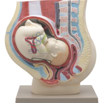
- Human Model – Pelvis with Baby: A pelvis with a fetal doll for practising engagement, station and rotation.
- Human Model – Delivery Doll: A delivery doll to pass through the pelvis when teaching the movements of labour.
- Human Adult Male Pelvis Structural Model: A male pelvis for comparing the inlet shape and subpubic angle with the female pelvis.
- Human Adult Female Pelvis Structural Model: Another female pelvis structural model showing the sacroiliac joints and pubic symphysis.
Frequently Asked Questions
What are the inlet, cavity and outlet of the female pelvis?
The inlet is the opening at the pelvic brim, where the baby’s head enters. The cavity is the curved passage below it. The outlet is the lower opening, bounded by the pubic arch in front, the ischial tuberosities at the sides and the coccyx behind.
What is a gynaecoid pelvis?
It is the typical female pelvis shape, with a round inlet, straight side walls, a wide subpubic arch and blunt ischial spines. This shape gives the most room for birth. Other shapes are described as android, anthropoid and platypelloid, although many real pelves fall between types.
What is the obstetric conjugate?
It is the shortest front-to-back distance the baby’s head must pass through at the inlet, measured from the sacral promontory to the nearest point on the back of the pubic symphysis. It cannot be measured directly in a woman, but on a pelvis model students can see exactly where it lies.
Why are the ischial spines important landmarks in labour?
They project into the middle of the pelvic cavity and can be felt during a vaginal examination. The level of the baby’s head is measured against them to give the station, and they mark the narrowest part of the cavity that the head must pass.
Can a fetal doll be passed through a female pelvis model?
Yes, a female pelvis is often used with a fetal doll or fetal skull to show how the head enters the inlet sideways, rotates in the cavity and leaves the outlet facing backwards. This is the core practical for teaching the mechanism of normal labour.
How is a female pelvis model used outside obstetrics?
It is used in anatomy and physiotherapy teaching to study the hip bones, sacroiliac joints and pubic symphysis, and the attachments of the hip and pelvic floor muscles. In biology it is a clear example of how skeleton shape relates to function.
Last Updated: November 2026








Reviews
There are no reviews yet.